iPSC-Derived Neurons Mirror LOAD Pathologies of Their Donors
Quick Links
Alzheimer’s researchers are making more use than ever of human induced pluripotent stem cell lines to model the disease. But how well do iPSCs generated from people with late-onset AD really reflect brain biology? In the August 26 Neuron online, researchers led by Tracy Young-Pearse at Brigham and Women’s Hospital, Boston, report that neurons made from 53 such lines exhibited gene-expression profiles similar to those of neurons in the postmortem brain of the corresponding iPSC donor. Strikingly, amyloidogenic APP processing in these neuronal cultures correlated with the plaque load in the donor’s brain at death, as well as with his or her rate of cognitive decline.
- High Aβ42 production in induced neurons reflected plaque load in the donor brain.
- Likewise, aggregated phospho-tau correlated with tangles.
- In the cultured neurons, protein phosphatase 1 activity linked Aβ42 to p-tau.
“Going into this study, we didn’t think we would see that kind of congruence. There are so many differences between cells in a dish and the brain,” Young-Pearse said. “But we were able to capture the genetically encoded risk of AD in neurons.”
The data also point toward specific pathways and proteins associated with pathological Aβ and tau. Young-Pearse believes these iPSC lines, which are freely available to scientists, will be useful for identifying disease mechanisms and testing therapeutics.
Jerome Mertens at the Salk Institute in La Jolla, California, agreed. “I am very thankful to the authors for providing such an outstandingly useful resource to the field,” he wrote to Alzforum. “This was a humongous tour de force.” (See full comment below.)
Michael Ward at the National Institute of Neurological Disorders and Stroke in Bethesda, Maryland, praised the study’s innovative approach. “This is a creative application of iPSC technologies, but also showcases some of the challenges in cellular modeling of sporadic neurodegenerative diseases,” Ward wrote (full comment below).
For example, he noted that the differences in APP processing between the AD and control groups were small, likely due to high genetic variability among individuals. Studying additional lines might strengthen the findings, Ward suggested.

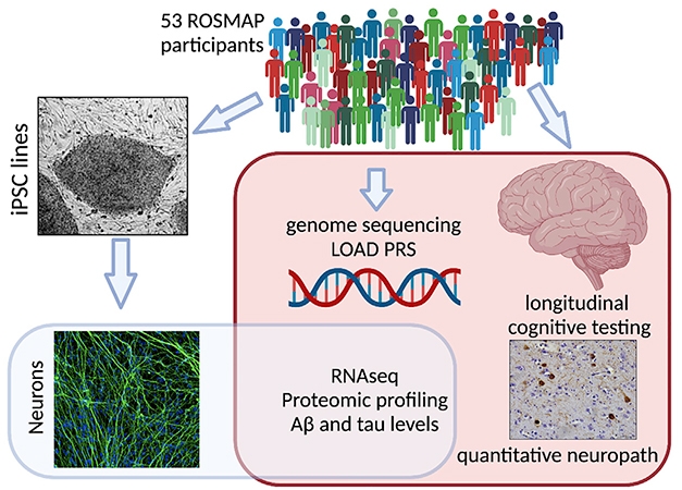
Data Mine. Genomic, proteomic, and transcriptomic phenotypes of induced neurons made from the ROSMAP cohort correlated with the neuropathological and cognitive status of their donors. [Courtesy of Lagomarsino et al., Neuron, 2021.]
Many of the Alzheimer’s iPSC lines studied so far have been generated from people with early onset AD, capturing strong amyloid phenotypes (Jan 2012 news; Apr 2019 news; Aug 2019 news). Although a few iPSC lines made from sporadic AD patients accumulated Aβ as well, no one had done a comprehensive study of how well such lines model AD (Feb 2013 news).
Joint first authors Valentina Lagomarsino and Richard Pearse II used cryopreserved peripheral blood mononuclear cells donated by 53 participants in the Religious Orders Study and Rush Memory and Aging Project (ROSMAP) to generate iPSC lines. All 53 donors had also agreed to donate their brains for autopsy, and have since died. Twenty of the brains had no Alzheimer’s pathology, 17 were amyloid-positive but the donor had been cognitively healthy at the time of death, and 16 came from people with a clinical and neuropathological AD diagnosis.
From these lines, the authors generated neuronal cultures that were 98 percent pure, with gene-expression profiles similar to those of cortical glutamatergic neurons. How closely would these immature, isolated cells resemble the mature neurons of the brains they came from?
The authors compared bulk RNA-Seq data from the induced neurons and from the medial prefrontal cortex of the corresponding donor’s brain. They found the gene-expression profiles were more alike than would be expected by chance, with about twice as many genes positively correlated as negatively correlated between the samples. This indicated that at least some of the neuronal gene expression seen in aging brain is genetically determined and can be modelled in culture.
The authors next identified subsets of genes that consistently varied between induced neurons made from AD patients and from cognitively normal people with either high or low amyloid pathology. They found more than 600 genes altered between the AD and low pathology groups, and nearly 300 between AD and high pathology. These expression changes fell into particular pathways. For example, neurons made from AD patients boosted expression of genes related to synaptic transmission and microtubule regulation, and suppressed those involved in protein production and apoptosis. Many of these differences replicated in the corresponding postmortem brain samples. A proteomic analysis of induced neurons and postmortem brain also identified these AD-related changes.
Importantly, the authors found that APP processing in induced neurons mirrored the AD status of the donor brain. Although the absolute amount of Aβ was not different between cultures, the higher the Aβ42/Aβ37 ratio in the culture media, the more plaques the corresponding brain had. This suggests that a shift in γ-secretase cleavage in favor of longer, more amyloidogenic peptides had promoted plaque deposition. That implies that increased Aβ42 production contributes to late-onset AD, just as it does to early onset.
A high Aβ42/Aβ37 ratio correlated with steeper cognitive decline as well. Young-Pearse believes this is not all just about Aβ42, but that Aβ37 may be protective, noting that the more of it induced neurons made, the better the donor’s cognition. Supporting this, a previous study had flagged Aβ42/Aβ37 as a stronger marker for AD than Aβ42/Aβ40 (Liu et al., 2021).
For tau, too, the authors found a relationship between induced neurons and brain. Detecting tau in cell lysates by western blot, they found that the amount of low-molecular-weight (LMW), monomeric p-tau isolated from induced neurons correlated with the amount of LMW p-tau in the corresponding brain sample. This p-tau reacted with antibodies to both p-tau181 and p-tau217.
Curiously, the more LMW p-tau a brain contained, the less high-molecular-weight (HMW), presumably aggregated, p-tau and fewer tangles it had. HMW tau in human brain has been reported to contain tau seeds and associate with more aggressive disease (Dujardin et al., 2020). Thus, an excess of LMW p-tau may reflect a relative scarcity of aggregated tau seeds.
In keeping with this, LMW p-tau in induced neurons correlated with better cognition in their donors, and HMW p-tau with worse. In fact, the type of p-tau in an induced neuron culture predicted the donor's cognitive status as well as or better than a person’s plaque and tangle burden did.

Missing Link? Protein phosphatase 1 (PP1) lies downstream of Aβ42 production and upstream of tau aggregation, potentially revealing a mechanistic connection between the pathologies. [Courtesy of Lagomarsino et al., Neuron, 2021.]
Altogether, the findings suggested that the genome of the donor exerts a strong influence on his or her late-onset AD risk. For each of them, the authors calculated a polygenic risk score based on GWAS hits, even those of nominal significance. A high PRS score correlated with the Aβ42/Aβ37 ratio in induced neurons, though not with p-tau.
This strengthened the idea that genetic risk in AD originates largely from APP processing and metabolism. High PRS also correlated with low levels of certain proteins, including the AD risk factor PICALM and protein phosphatase 1.
PP1 drew the authors’ interest because it dephosphorylates tau and is low in AD brain. Following up, the scientists found that PP1 activity was associated with a low Aβ42/Aβ37 ratio in both induced neurons and brain. Boosting the Aβ42/Aβ37 ratio in induced neurons by tweaking APP processing lowered PP1 activity and increased HMW p-tau. On the other hand, pharmacologically inhibiting PP1 in induced neurons boosted HMW p-tau, but had no effect on Aβ42/Aβ37. Together, the findings hint that PP1 alterations occur downstream of Aβ changes and upstream of tau, and may help link the two pathologies. PP1 has long been studied in AD (e.g., Sun et al., 2003).
Despite the PRS correlations, induced neurons likely miss much of the genetic risk of AD, which is now believed to be borne in large part by microglia (Jun 2017 news; Apr 2019 conference news; Aug 2019 news). In ongoing work, Young-Pearse is examining Aβ clearance in microglia and astrocytes generated from the same PBMC-derived iPSC lines.
Another limitation is that induced neurons are immature, making them poor models for aging brain. Recent work suggests that neurons generated directly from human fibroblasts, without going through an iPSC stage, retain epigenetic marks of aging and might better model brain diseases (Jun 2021 news). Young-Pearse noted that this approach is an option in the ROSMAP cohort as well, because fibroblasts from the participants are available. However, fibroblast cultures cannot be expanded and shared as readily as immortal iPSC lines, which is why many labs are sticking to the latter for the time being.
On that note, the iPSC Neurodegenerative Disease Initiative, co-led by Ward, uses isogenic iPSC lines to investigate the effects of specific AD mutations (May 2021 news). Young-Pearse said this strategy complements her sporadic AD lines, which represent more of a “Wild West” of diverse genetic expression. She believes both approaches will help dissect AD mechanisms.
Several labs are already using Young-Pearse’s lines to investigate the connection between diabetes and AD, and how viral and bacterial infections affect pathogenesis. Because of the rich data available from the ROSMAP cohort, which includes full genome sequencing on every participant, the lines are well-suited to explore links between gene expression and health outcomes, Young-Pearse said.—Madolyn Bowman Rogers
References
News Citations
- Induced Neurons From AD Patients Hint at Disease Mechanisms
- Familial Alzheimer’s Mutations: Different Mechanisms, Same End Result
- Familial AD Mutations, β-CTF, Spell Trouble for Endosomes
- Aβ Oligomers Linked to ER Stress in Patient-Derived Neurons
- Microglial Master Regulator Tunes AD Risk Gene Expression, Age of Onset
- Parsing How Alzheimer’s Genetic Risk Works Through Microglia
- AD Genetic Risk Tied to Changes in Microglial Gene Expression
- Neurons Made from Fibroblasts Keep Imprint of Alzheimer's, Aging
- iNDI Aims to Standardize Human Stem Cell Research
Paper Citations
- Liu L, Lauro BM, Wolfe MS, Selkoe DJ. Hydrophilic loop 1 of Presenilin-1 and the APP GxxxG transmembrane motif regulate γ-secretase function in generating Alzheimer-causing Aβ peptides. J Biol Chem. 2021;296:100393. Epub 2021 Feb 8 PubMed.
- Dujardin S, Commins C, Lathuiliere A, Beerepoot P, Fernandes AR, Kamath TV, De Los Santos MB, Klickstein N, Corjuc DL, Corjuc BT, Dooley PM, Viode A, Oakley DH, Moore BD, Mullin K, Jean-Gilles D, Clark R, Atchison K, Moore R, Chibnik LB, Tanzi RE, Frosch MP, Serrano-Pozo A, Elwood F, Steen JA, Kennedy ME, Hyman BT. Tau molecular diversity contributes to clinical heterogeneity in Alzheimer's disease. Nat Med. 2020 Aug;26(8):1256-1263. Epub 2020 Jun 22 PubMed. Correction.
- Sun L, Liu SY, Zhou XW, Wang XC, Liu R, Wang Q, Wang JZ. Inhibition of protein phosphatase 2A- and protein phosphatase 1-induced tau hyperphosphorylation and impairment of spatial memory retention in rats. Neuroscience. 2003;118(4):1175-82. PubMed.
Further Reading
News
- Expression, Expression, Expression—Time to Get on Board with eQTLs
- Umbilical Cord With Presenilin Mutation Births New Cell Model of Familial AD
- Introducing: iPSC Collection from Tauopathy Patients
- Reproducible Brain Organoids Could Offer New Models for Research
- iPSC Disease Models Up and Coming for AD, Down’s, ALS
Primary Papers
- Lagomarsino VN, Pearse RV 2nd, Liu L, Hsieh YC, Fernandez MA, Vinton EA, Paull D, Felsky D, Tasaki S, Gaiteri C, Vardarajan B, Lee H, Muratore CR, Benoit CR, Chou V, Fancher SB, He A, Merchant JP, Duong DM, Martinez H, Zhou M, Bah F, Vicent MA, Stricker JM, Xu J, Dammer EB, Levey AI, Chibnik LB, Menon V, Seyfried NT, De Jager PL, Noggle S, Selkoe DJ, Bennett DA, Young-Pearse TL. Stem cell-derived neurons reflect features of protein networks, neuropathology, and cognitive outcome of their aged human donors. Neuron. 2021 Nov 3;109(21):3402-3420.e9. Epub 2021 Sep 1 PubMed.
Annotate
To make an annotation you must Login or Register.

Comments
National Institutes of Health
This is a creative application of iPSC technologies, but also showcases some of the challenges in cellular modeling of sporadic neurodegenerative diseases. The authors performed an innovative cross-analysis of data from iPSC-derived neurons and matched postmortem brain from healthy and AD individuals, identifying potential dysregulated pathways in AD. Rich clinical phenotyping was performed on all of the iPSC donors as part of the ROS and MAP cohorts, adding further value to the published datasets.
However, as the authors note, the effect sizes observed in the iPSC neuron experiments were quite small, despite the use of robust NGN2-based differentiation methods. This is potentially due to multiple factors, including inherent variability across iPSC lines and genetic differences among donors, coupled with the (likely) divergent biology of sporadic AD and relatively modest sample sizes (n=16 LOAD).
It will be interesting to see whether iPSC-based studies of other sporadic neurodegenerative diseases—such as those being undertaken by Answer ALS—are able to overcome these challenges by employing substantially higher sample sizes of greater than 1,000 participants. In the future, it will also be interesting to directly compare lines that harbor familial mutations and lines from individuals with sporadic disease.
Neural Aging Lab, University of Innsbruck
Reading this paper in Neuron felt extremely real on many levels. I am thankful to the authors for providing such an outstandingly useful resource to the field. It will accelerate discovery in the Alzheimer's field.
This was a humongous tour de force, not only for the deep multi-omic and functional phenotyping work, but also for the clear, logical, honest data analysis that takes into account a realistic representation of individual variability in pathology, cognition, and iPSC neuronal phenotypes.
I was pleasantly surprised to see that the authors were able to find a significant association between amyloid and tau in iPSC neurons and the cognitive trajectories in the same individuals. These data suggest that, at least for a subset of "high pathology" patients, established disease pathways are linked to genetic risk, and can be used as in vitro readouts also for sporadic LOAD models. I was not necessarily expecting this.
Johns Hopkins University
Really nice study. Similarly, we found in a cohort of about 35 sporadic ALS iPS cell lines and C9ORF72 iPS neuronal lines, derived from PBMC and not fibroblasts, that the protein and pathophysiological events very closely mirrored that found in autopsy cortex from sporadic and C9ORF72 ALS patients (Coyne et al., 2020; Coyne et al., 2021).
There is no question that iPS cells can serve as a foundational tool to unravel cell biological events in these diseases. However, one needs to look at larger number of iPS lines, no different than examining multiple patients. Studies in a few iPS lines, especially for sporadic disorders, require large number of lines—not a handful—to be scientifically meaningful.
Although some argue the need to use fibroblasts as a starting point, clearly, emerging and robust data suggest that blood-derived iPS lines can very closely mirror the actual cell biological defect found in authentic human autopsy materials.
References:
Coyne AN, Zaepfel BL, Hayes L, Fitchman B, Salzberg Y, Luo EC, Bowen K, Trost H, Aigner S, Rigo F, Yeo GW, Harel A, Svendsen CN, Sareen D, Rothstein JD. G4C2 Repeat RNA Initiates a POM121-Mediated Reduction in Specific Nucleoporins in C9orf72 ALS/FTD. Neuron. 2020 Sep 23;107(6):1124-1140.e11. Epub 2020 Jul 15 PubMed.
Coyne AN, Baskerville V, Zaepfel BL, Dickson DW, Rigo F, Bennett F, Lusk CP, Rothstein JD. Nuclear accumulation of CHMP7 initiates nuclear pore complex injury and subsequent TDP-43 dysfunction in sporadic and familial ALS. Sci Transl Med. 2021 Jul 28;13(604) PubMed.
Make a Comment
To make a comment you must login or register.