Organoids Implicate Cholesterol Dysregulation in Tau Pathology
Quick Links
When studying neurodegenerative disease in human tissue, scientists often have trouble pinning down what went wrong first, since they must usually look at postmortem brain samples. Newer research techniques, such as the generation of cerebral organoids from human stem cells, are helping parse out the initial steps on the pathway to disease. Case in point: In the September 13 Stem Cell Reports, researchers led by Kenneth Kosik at the University of California, Santa Barbara, in collaboration with Sally Temple at the Neural Stem Cell Institute of Rensselaer, New York, describe the use of organoids to study the effect of pathogenic tau mutations. Over the first few months in culture, the main effect of these mutations was to boost cholesterol production by astrocytes. Though the researchers do not know the mechanisms involved, the data fit with a growing body of research showing disruption of lipid metabolism in neurodegenerative disease.
- Human cerebral organoids allow the study of neuron-astrocyte interactions.
- Mutant tau, which is mainly neuronal, increases cholesterol production in astrocytes.
- Is this boost compensatory or harmful?
The findings may point toward new research directions, Kosik believes. “This shows it is possible to use human brain organoids to study a human neurodegenerative disease,” he told Alzforum. This, even though organoids contain many more progenitor and newborn cells than do older human brains.
Julia TCW at Boston University noted that most studies of tauopathy have focused on neurons. She believes the new findings of dysregulated cholesterol in astrocytes are significant and could suggest new therapeutic targets for early intervention.

Cholesterol Boom. Astrocytes (red) carrying mutant tau (bottom) make much more HMGCS1 (green), an enzyme in the cholesterol biosynthesis pathway, compared to cells with wild-type tau (top). [Courtesy of Glasauer et al., Stem Cell Reports.]
Protocols for generating cerebral organoids have been around for a decade, with recent technical advances making them more reproducible and reliable (Aug 2013 news; Jun 2019 news). Kosik and colleagues employed a protocol developed at Stanford University that produces long-lived spherical organoids enriched for pyramidal cortical neurons and astrocytes (Yoon et al., 2019).
First author Stella Glasauer used induced pluripotent stem cells made from three people with heterozygous V337M tau mutations, two with heterozygous R406W, and one with homozygous R406W to generate dozens of cerebral organoids. In people, these mutations cause frontotemporal dementia. By three months in culture, the organoids consisted of mostly excitatory pyramidal cells, with smaller populations of astrocytes and inhibitory interneurons. Glasauer and colleagues then isolated cells from pooled samples of three to four genetically identical organoids for single-cell RNA-Seq. They compared tau mutant organoids with genetically corrected isogenic controls at several different ages. They analyzed around 1,200 cells per sample, for a total of 76,111 cells.
In excitatory neurons with mutant tau, 60 genes went down and 81 genes up compared with isogenic controls. Suppressed genes were mostly associated with glycolysis, the biochemical process that produces energy in the absence of oxygen, while several of the elevated genes were mitochondrial. In inhibitory neurons, GABA receptors and glycolytic genes were suppressed.

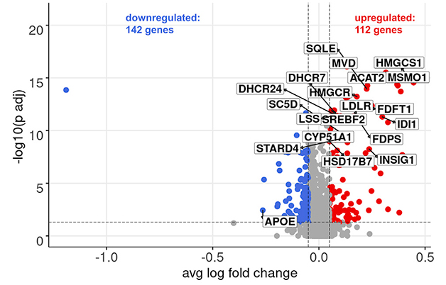
Lipids Gone Wild. Most gene expression changes in human cerebral organoids carrying mutant tau were in astrocytes, where 142 genes were down (blue dots) and 112 up (red dots) compared to controls organoids; in particular, multiple genes responsible for cholesterol metabolism and fatty acid synthesis were highly elevated (boxes). [Courtesy of Glasauer et al., Stem Cell Reports.]
However, most changes were in astrocytes, where 142 genes were down and 112 up (see image above). In particular, numerous genes involved in cholesterol synthesis, metabolism, and transport were highly expressed in astrocytes with mutant tau. These changes were more pronounced in cells with homozygous than heterozygous R406W. They also became more dramatic with age, with cholesterol synthesis genes more elevated in 8-month-old organoids than in 4-month-olds. In addition, genes responsible for fatty acid synthesis went up.
Curiously, APOE expression was low in astrocytes with mutant tau. This was true regardless of whether they carried APOE2, 3, or 4. Some previous studies have linked APOE4 to disrupted lipid metabolism in astrocytes, but this was in the context of Alzheimer’s disease, in the presence of wild-type tau (Apr 2019 conference news; Aug 2019 news; Nov 2021 conference news).
The authors verified some of these RNA-Seq findings at the lipid level, using liquid chromatography mass spectrometry to identify several cholesterol pathway components in organoids and isogenic controls made from three of the cell lines. They detected high levels of cholesterol, three of its precursors, and one metabolite, in 7-month-old mutant tau organoids, but not in 4-month-old. This suggested that expression changes do affect lipid synthesis, but with a time delay, since the transcripts were already elevated at four months.
How does mutant tau juice cholesterol metabolism? While this is unknown, Kosik noted that neurons need high amounts of cholesterol in their plasma membrane, and this is produced by astrocytes. He speculated that mutant tau oligomers might damage neuronal membranes, provoking astrocytes to ramp up cholesterol production to compensate and repair the damage. A previous study found that cholesterol in neuronal membranes helps keep out tau aggregates, protecting cells (May 2022 news). Conversely, it is equally possible that elevated cholesterol is harmful, since its immediate metabolites, cholesterol esters, have been shown to be neurotoxic and promote tau phosphorylation (Feb 2019 news).
In future work, Kosik will further characterize the phenotype of mutant tau organoids, particularly their lipid composition, to figure out whether these changes are harmful or helpful. He will also look for early signs of tau pathology, such as hyperphosphorylation or aggregation. A previous study of V337M cerebral organoids reported hyperphosphorylated tau and loss of excitatory neurons by six months in culture, suggesting these structures can reproduce some of the features of FTD (Bowles et al., 2021).—Madolyn Bowman Rogers
References
News Citations
- Mini Brain in a Dish Models Human Development
- Reproducible Brain Organoids Could Offer New Models for Research
- Could Greasing the Wheels of Lipid Processing Treat Alzheimer’s?
- ApoE4 Glia Bungle Lipid Processing, Mess with the Matrisome
- Do Lipids Lubricate ApoE's Part in Alzheimer Mechanisms?
- Membrane Border Patrol: Cholesterol Stymies Tau Uptake, Aggregation
- Cholesteryl Esters Hobble Proteasomes, Increase p-Tau
Paper Citations
- Yoon SJ, Elahi LS, Pașca AM, Marton RM, Gordon A, Revah O, Miura Y, Walczak EM, Holdgate GM, Fan HC, Huguenard JR, Geschwind DH, Pașca SP. Reliability of human cortical organoid generation. Nat Methods. 2019 Jan;16(1):75-78. Epub 2018 Dec 20 PubMed.
- Bowles KR, Silva MC, Whitney K, Bertucci T, Berlind JE, Lai JD, Garza JC, Boles NC, Mahali S, Strang KH, Marsh JA, Chen C, Pugh DA, Liu Y, Gordon RE, Goderie SK, Chowdhury R, Lotz S, Lane K, Crary JF, Haggarty SJ, Karch CM, Ichida JK, Goate AM, Temple S. ELAVL4, splicing, and glutamatergic dysfunction precede neuron loss in MAPT mutation cerebral organoids. Cell. 2021 Aug 19;184(17):4547-4563.e17. Epub 2021 Jul 26 PubMed.
Further Reading
Primary Papers
- Glasauer SM, Goderie SK, Rauch JN, Guzman E, Audouard M, Bertucci T, Joy S, Rommelfanger E, Luna G, Keane-Rivera E, Lotz S, Borden S, Armando AM, Quehenberger O, Temple S, Kosik KS. Human tau mutations in cerebral organoids induce a progressive dyshomeostasis of cholesterol. Stem Cell Reports. 2022 Sep 13;17(9):2127-2140. Epub 2022 Aug 18 PubMed.
Annotate
To make an annotation you must Login or Register.

Comments
Boston University School of Medicine
Studies on tauopathy, especially those of MAPT mutations, have mainly focused on neurons, since MAPT is expressed in these cells and is known to dysregulate them. This study, using human organoids composed of various neurons and glia, comes from an interesting angle. The astrocytes express both mutant and control MAPT in the context of a brain environment that includes multiple cell types, and the findings suggest both a cell-autonomous effect of MAPT in astrocytes and a non-cell-autonomous effect in mutant astrocytes driven from mutant MAPT in neurons. Regardless, they report that a major defect in tauopathy mutants in astrocytes was upregulation of cholesterol biosynthesis, which we have reported as the major dysregulation caused by APOE4 in glia in AD and in many other neurodegenerative diseases (TCW et al., 2022). The findings suggest a potential therapeutic target for early intervention.…More
References:
Tcw J, Qian L, Pipalia NH, Chao MJ, Liang SA, Shi Y, Jain BR, Bertelsen SE, Kapoor M, Marcora E, Sikora E, Andrews EJ, Martini AC, Karch CM, Head E, Holtzman DM, Zhang B, Wang M, Maxfield FR, Poon WW, Goate AM. Cholesterol and matrisome pathways dysregulated in astrocytes and microglia. Cell. 2022 Jun 23;185(13):2213-2233.e25. PubMed. BioRxiv.
Dartmouth Medical School
Dartmouth Medical School
We read the work of Glasauer et al. with great interest. The authors performed scRNA-Seq, immunohistochemical staining, and lipidomic analyses in human organoids. They elegantly demonstrated that mutant taus upregulated the expressions of several enzymes in the cholesterol biosynthetic pathway, including HMG-CoA synthase and HMG-CoA reductase, in astrocytes. Thus, upregulation in cholesterol biosynthesis in astrocytes is one of the significant responses to the pathogenic cascade initiated by mutant forms of tau.…More
The authors found that ACAT2 expression in astrocytes is also upregulated. On page 2131, they wrote, "ACAT2, encoding an enzyme converting cholesterol to its storage form cholesteryl esters." We feel it is important to point out that in this work, ACAT2 does not stand for a cholesterol storage enzyme; instead, it stands for acetyl-CoA C-acetyltransferase, which is the first enzyme in the cholesterol biosynthetic pathway. As an off-topic comment: ACAT1 stands for a distinct acetyl-CoA acetyltransferase located in the mitochondria and plays a crucial role in ketogenesis.
The enzyme responsible for converting cholesterol to its storage form is known as "acyl-CoA: cholesterol acyltransferase." For many years, it has been abbreviated as ACAT. There are two ACATs. ACAT1 was identified in 1993; ACAT2 was identified in 1998. To avoid confusion with acetyl-CoA acetyltransferases, the HUGO Gene Nomenclature Committee recommended that acyl-CoA: cholesterol acyltransferase be renamed sterol O-acyltransferases and abbreviated as SOATs. In recent years, these enzymes are often referred to as ACATs/SOATs.
Both ACAT1/SOAT1 and ACAT2/SOAT2 are multi-span membrane proteins located at the endoplasmic reticulum. Unlike enzymes in the cholesterol biosynthetic pathway, neither ACAT1/SOAT1 nor ACAT2/SOAT2 is transcriptionally regulated by the master transcription factor SREBP2. Instead, each enzyme is allosterically activated by sterols, including cholesterol and oxysterols; this mechanism enables ACATs/SOATs to respond rapidly to changes in cellular sterol levels. ACATs/SOATs are targeted to treat several human diseases, including cardiovascular disease, neurodegenerative diseases, and certain forms of cancer. A recent review on ACATs/SOATs is available (Rogers et al., 2015).
—Michael Duong of University of Pennsylvania School of Medicine co-authored this comment.
References:
Rogers MA, Liu J, Song BL, Li BL, Chang CC, Chang TY. Acyl-CoA:cholesterol acyltransferases (ACATs/SOATs): Enzymes with multiple sterols as substrates and as activators. J Steroid Biochem Mol Biol. 2015 Jul;151:102-7. Epub 2014 Sep 12 PubMed.
View all comments by Catherine ChangUniversity of California, Santa Barbara
University of California, Santa Barbara
We would like to thank Drs. Ta-Yuan Chang and Catherine Chang, and Michael Duong for their interest in our paper and their comment.
The identifier ACAT has been used ambiguously in the literature: acyl-CoA: cholesterol acyltransferases (catalyzing esterification of cholesterol) and acetyl-CoA acetyltransferases (catalyzing the conversion of two units of acetyl-CoA to acetoacetyl-CoA) have both been referred to as ACATs, even in the recent literature.…More
The comment’s authors correctly state that the gene symbol ACAT2, which we refer to in our paper, encodes an acetyl-CoA acetyltransferase according to current gene name convention.
This does not change the story of our paper and will only change two sentences in the results section.
Specifically, currently we write:
15 of these genes encode enzymes of the cholesterol synthesis pathway, including its rate limiting enzyme HMGCR (Fig. 3A). Also ACAT2, encoding an enzyme converting cholesterol to its storage form cholesteryl esters, STARD4, encoding an intracellular cholesterol transporter, LDLR, encoding an important receptor for cholesterol uptake, and SREBF2, encoding a transcriptional activator of cholesterol biosynthesis enzymes, were significantly upregulated (Figure 3A).
Instead, it should say:
16 of these genes encode enzymes of the cholesterol synthesis pathway, including its rate limiting enzyme HMGCR (Fig. 3A). Also STARD4, encoding an intracellular cholesterol transporter, LDLR, encoding an important receptor for cholesterol uptake, and SREBF2, encoding a transcriptional activator of cholesterol biosynthesis enzymes, were significantly upregulated (Figure 3A).
View all comments by Kenneth KosikAmsterdam UMC, loc. VUmc
Maastricht University; VU University Medical Centre
In this work, Glasauer and colleagues investigated RNA expression differences of pyramidal neurons and astrocytes from organoids with a MAPT mutation compared to their isogenic controls. Most differences were found in neurons, where MAPT also has the highest expression. Sixty genes were downregulated and associated with glucose metabolism, 80 genes were upregulated and associated with mitochondrial function.…More
Glasauer and colleagues next compared astrocytes between MAPT mutation and control organoids, and observed upregulated genes associated with cholesterol metabolism, including SREBF2. SREBF2 is a transcription factor that regulates cholesterol metabolism. Another study recently also identified SREBF2 to be implicated in cholesterol metabolism alterations that were associated with the APOE e4 genotype (TCW et al., 2022). SREBF2 can be activated by endoplasmic reticulum stress, and can subsequently disrupt lipid metabolism (Colgan et al., 2007).
In Alzheimer’s disease and other tauopathies, aggregation of tau can cause endoplasmic reticulum stress (Hoozemans and Scheper, 2012). It would be interesting to further study the role of phosphorylated tau in endoplasmic reticulum stress and cholesterol metabolism in the MAPT organoids.
References:
Tcw J, Qian L, Pipalia NH, Chao MJ, Liang SA, Shi Y, Jain BR, Bertelsen SE, Kapoor M, Marcora E, Sikora E, Andrews EJ, Martini AC, Karch CM, Head E, Holtzman DM, Zhang B, Wang M, Maxfield FR, Poon WW, Goate AM. Cholesterol and matrisome pathways dysregulated in astrocytes and microglia. Cell. 2022 Jun 23;185(13):2213-2233.e25. PubMed. BioRxiv.
Colgan SM, Tang D, Werstuck GH, Austin RC. Endoplasmic reticulum stress causes the activation of sterol regulatory element binding protein-2. Int J Biochem Cell Biol. 2007;39(10):1843-51. Epub 2007 May 16 PubMed.
Hoozemans JJ, Scheper W. Endoplasmic reticulum: The unfolded protein response is tangled in neurodegeneration. Int J Biochem Cell Biol. 2012 Aug;44(8):1295-8. PubMed.
View all comments by Pieter Jelle VisserMake a Comment
To make a comment you must login or register.