Sylvain Lesné, Who Found Aβ*56, Accused of Image Manipulation
Quick Links
The Alzheimer’s field was rocked this week by allegations against Sylvain Lesné at the University of Minnesota, Minneapolis. Lesné stands accused of manipulating data images in multiple papers, including his 2006 Nature paper identifying Aβ*56 as a toxic oligomer associated with cognitive decline. The potentially altered images were found by neuroscientist Matthew Schrag at Vanderbilt University. Earlier this year, Schrag alerted the National Institutes of Health and UMN, as well as the journals that published the papers. Multiple investigations are ongoing.
A July 21 Science article by investigative journalist Charles Piller broke the news to the field at large. Science magazine conducted its own six-month investigation, in which independent analysts agreed the images showed signs of tampering.
Alzheimer's researchers expressed dismay. Most thought that even if the allegations are confirmed, the impact on oligomer research would be much smaller than the general effect of bringing disrepute to the field. “The refutation of Aβ*56 would have no impact on the huge weight of evidence that supports a role for soluble aggregates (aka oligomers) in AD,” Dominic Walsh at Brigham and Women’s Hospital, Boston, wrote to Alzforum. Mathias Jucker at the University of Tübingen, Germany, concurred. “The Aβ*56 work was just one paper among many others claiming that Aβ oligomers are key toxic species in AD pathogenesis. I do not think the field would have developed differently without the Lesné work,” he wrote.
“I am speechless about these allegations. This damages the reputation of the oligomer research field, where much good work is being done,” Christian Haass of DZNE Munich wrote to Alzforum (full comments below).
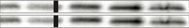
Copies? In Lesné’s 2006 Nature paper on Aβ*56, these western blot bands ostensibly represent two different control proteins, but instead are exact duplicates of each other. [Courtesy of Science/AAAS.]
Lesné identified Aβ*56 while in Karen Ashe’s lab at UMN. The finding made a splash at the time, generating excitement as a potential link between a specific Aβ oligomer and cognitive decline (Mar 2006 news). However, few if any subsequent papers have been published on it outside of Ashe and Lesné’s labs. Likewise, other scientists working on aggregated Aβ wrote that they were unaware of independent corroboration (comments below).
Many Alzheimer’s researchers told Alzforum, some off the record, that they tried but were unable to replicate the findings. Most did not publish those efforts. One who did was Dennis Selkoe at Brigham and Women’s. At the time, Selkoe reported being unable to find the species in both human cortical extracts and cerebrospinal fluid (Shankar et al., 2008; Klyubin et al., 2008).
Schrag became aware of potential problems with Lesné’s work while being contracted to investigate an entirely separate issue, namely allegations against biotech Cassava Sciences over its drug simufilam. In the course of that work, Schrag perused PubPeer, an online site where researchers flag suspected problems in published work. Schrag spotted complaints about figures in Lesné’s work. Digging deeper, he flagged figures in 20 Lesné papers; 10 of which involved Aβ*56. The problems included duplicated bands on western blots (see image above), as well as images that seemed to be composites from different experiments, or figures reprinted in later papers as though new. Lesné did not respond to a request for comment from Alzforum.
Schrag found no suspicious figures in papers from the Ashe lab where Lesné was not a co-author. Ashe is not under investigation.
Schrag submitted his concerns to NIH in January 2022 and alerted the journals in question. In response, at least two journals, Nature and Science Signaling, have published “expressions of concern” about the papers, and are investigating. UMN also says it is reviewing the matter. NIH whistleblower complaints are typically referred to the Office of Research Integrity; investigations there can take years.
Schrag also contacted Science. The journal showed the data to two independent image analysts, Elisabeth Bik and Jana Christopher, as well as to Alzheimer’s researchers including Selkoe; George Perry at the University of Texas, San Antonio; Donna Wilcock at the University of Kentucky, Lexington; and John Forsayeth at the University of California, San Francisco. All agreed there were genuine issues with the figures.
Piller also uncovered previous suspicions about Lesné’s work. His postdoc supervisor Denis Vivien at the University of Caen Normandy, France, told Science that he withdrew a manuscript he had been co-authoring with Lesné over doubts about some immunostainings, and that others in his lab were unable to replicate them.
Ashe declined to comment on the allegations against Lesné, but stands by the science behind Aβ*56. “Staff scientists in our labs regularly and reproducibly detect Aβ*56 in a subset of Tg2576 and J20 mice,” she wrote to Alzforum (full comment below).
Ashe's group initially reported finding the species in human CSF; however, a subsequent paper suggested that could have been an artifact, with the bands perhaps being confounded by N-terminal amyloid precursor protein fragments (Mar 2013 news; Grant et al., 2019). Another recent paper from the lab implied that the Aβ*56 bands in earlier publications could have been artifacts of using the biotin-avidin system and/or Protein A shed from Sepharose beads (Grant et al., 2019).
Whatever the effect of the allegations on Aβ*56 and oligomer research, AD researchers agreed they give the field a black eye. “This is not a real scientific problem, but it is most unfortunate for general science credibility,” Selkoe wrote to Alzforum.
Others noted that the scientific process tends to ferret out results that cannot be reproduced. “I am very disappointed to read this article of possible ‘fabrication’ in this field. But science is self-correcting, and this is a good example,” Colin Masters at the University of Melbourne, Australia, wrote to Alzforum.—Madolyn Bowman Rogers
References
News Citations
- Aβ Star is Born? Memory Loss in APP Mice Blamed on Oligomer
- Aβ*56 Found in Human CSF, Correlates With Tau?
Therapeutics Citations
Paper Citations
- Shankar GM, Li S, Mehta TH, Garcia-Munoz A, Shepardson NE, Smith I, Brett FM, Farrell MA, Rowan MJ, Lemere CA, Regan CM, Walsh DM, Sabatini BL, Selkoe DJ. Amyloid-beta protein dimers isolated directly from Alzheimer's brains impair synaptic plasticity and memory. Nat Med. 2008 Aug;14(8):837-42. PubMed.
- Klyubin I, Betts V, Welzel AT, Blennow K, Zetterberg H, Wallin A, Lemere CA, Cullen WK, Peng Y, Wisniewski T, Selkoe DJ, Anwyl R, Walsh DM, Rowan MJ. Amyloid beta protein dimer-containing human CSF disrupts synaptic plasticity: prevention by systemic passive immunization. J Neurosci. 2008 Apr 16;28(16):4231-7. PubMed.
- Grant MK, Handoko M, Rozga M, Brinkmalm G, Portelius E, Blennow K, Ashe KH, Zahs KR, Liu P. Human cerebrospinal fluid 6E10-immunoreactive protein species contain amyloid precursor protein fragments. PLoS One. 2019;14(2):e0212815. Epub 2019 Feb 28 PubMed.
- Grant MK, Shapiro SL, Ashe KH, Liu P, Zahs KR. A Cautionary Tale: Endogenous Biotinylated Proteins and Exogenously-Introduced Protein A Cause Antibody-Independent Artefacts in Western Blot Studies of Brain-Derived Proteins. Biol Proced Online. 2019;21:6. Epub 2019 Apr 18 PubMed.
Further Reading
Primary Papers
- Piller C. Blots on a field?. Science. 2022 Jul 22;377(6604):358-363. Epub 2022 Jul 21 PubMed.
Annotate
To make an annotation you must Login or Register.
Comments
University of Minnesota
Regarding Charles Piller’s article in Science, I cannot comment on the allegations about images that may have been inappropriately altered by my former co-worker Dr. Sylvain Lesné, because he is now under formal investigation at the University of Minnesota. However, I will comment on his scientific statements, because his description of my scientific thesis is inaccurate.
This Science article implied that my work has misled researchers in the Alzheimer’s field by encouraging the development of therapies targeting amyloid plaques, which most of us know are composed of Aβ. In fact, for over 20 years, I have consistently expressed concerns that drugs targeting plaques were likely to be ineffective. Based on my published work (Liu et al., 2015; Ashe, 2020), it is clear that there are two general forms of Aβ, type 1 and type 2. One particular form of type 1 (referred to in our papers as Aβ*56 and in the Science article as “toxic oligomers”) was shown by my lab and others to impair memory function in mice. The type 2 form of Aβ is the one found in amyloid plaques. It is this latter form that drug developers have repeatedly but unsuccessfully targeted. There have been no clinical trials targeting the type 1 form of Aβ, the form which my research has suggested is more relevant to dementia. Mr. Piller erroneously conflated the two forms of Aβ.
The readers of Mr. Piller’s article are given the strong impression that the pursuit of Aβ-targeted therapies for Alzheimer’s, which I agree has been frustratingly negative and expensive, was somehow ignited and/or fueled by the 2006 Nature paper. Readers must know that this is simply untrue and that decades of human genetics and murine models from many labs had led many drug developers to conclude that Aβ was a highly plausible target.
Mr. Piller’s article conflated two distinct issues: a) the frustrations regarding the difficulties of drug development in Alzheimer’s; and b) a specific accusation of scientific misconduct relating to a set of papers about one particular aspect of the Aβ hypothesis. To give the reader the impression that the latter bears a heavy burden of responsibility for the former, as he did in his article, is misleading.
Having worked for decades to understand the cause of Alzheimer disease, so that better treatments can be found for patients, it is devastating to discover that a co-worker may have misled me and the scientific community through the doctoring of images. It is, however, additionally distressing to find that a major scientific journal has flagrantly misrepresented the implications of my work.
For some additional scientific background, I would like to comment on the status of Aβ*56 and Aβ oligomer research more broadly. We previously reported that Aβ*56 is an SDS-stable Aβ assembly that impairs memory function in Tg2576 mice (Lesné et al., 2006). Dr. Peng Liu and other colleagues in my lab have gone on to confirm and extend these findings; we classified Aβ oligomers into two types, type 1 and type 2, that differ in structure, spatial distribution, and temporal expression, and hypothesized that type 1 are formed by primary nucleation and type 2 by secondary nucleation (Liu et al., 2015).
Based on these data, I hypothesize that Aβ*56 is a metastable type 1 oligomer, which can make it frustratingly elusive. Its detection must be conducted with care, as described in a paper by Ms. Marianne Grant, Dr. Kathy Zahs, and colleagues (Grant et al., 2019). [Editor’s note: Since 2017, Kathy Zahs has been a biocuration scientist at Alzforum].
Using canonical Aβ-epitope-specific antibodies, we have confirmed that a high-molecular-weight, SDS-stable Aβ assembly corresponds to Aβ*56 (Grant et al., 2019; Liu et al., manuscript in preparation). Dr. Liu and other staff scientists in our labs regularly and reproducibly detect Aβ*56 in a subset of Tg2576 and J20 mice. A major focus of our current work is to determine the secondary and quaternary biophysical structures of type 1 oligomers, which may help explain why current Aβ therapies which target type 2 oligomers may not have been as effective as once hoped. Another focus is to develop monoclonal antibodies against type 1 oligomers, which could pave a path forward toward new Aβ therapeutics.
References:
Lesné S, Koh MT, Kotilinek L, Kayed R, Glabe CG, Yang A, Gallagher M, Ashe KH. A specific amyloid-beta protein assembly in the brain impairs memory. Nature. 2006 Mar 16;440(7082):352-7. PubMed.
Ashe KH. The biogenesis and biology of amyloid β oligomers in the brain. Alzheimers Dement. 2020 Nov;16(11):1561-1567. Epub 2020 Jun 16 PubMed. Correction.
Liu P, Reed MN, Kotilinek LA, Grant MK, Forster CL, Qiang W, Shapiro SL, Reichl JH, Chiang AC, Jankowsky JL, Wilmot CM, Cleary JP, Zahs KR, Ashe KH. Quaternary Structure Defines a Large Class of Amyloid-β Oligomers Neutralized by Sequestration. Cell Rep. 2015 Jun 23;11(11):1760-71. Epub 2015 Jun 4 PubMed.
Grant MK, Shapiro SL, Ashe KH, Liu P, Zahs KR. A Cautionary Tale: Endogenous Biotinylated Proteins and Exogenously-Introduced Protein A Cause Antibody-Independent Artefacts in Western Blot Studies of Brain-Derived Proteins. Biol Proced Online. 2019;21:6. Epub 2019 Apr 18 PubMed.
Grant MK, Handoko M, Rozga M, Brinkmalm G, Portelius E, Blennow K, Ashe KH, Zahs KR, Liu P. Human cerebrospinal fluid 6E10-immunoreactive protein species contain amyloid precursor protein fragments. PLoS One. 2019;14(2):e0212815. Epub 2019 Feb 28 PubMed.
The University of Queensland
This is very sad to hear. I am a co-author on one of the papers in question (Amar et al., 2017). Sylvain had reached out to me at the time, as we had just made tau-isoform-specific antibodies. As a collaborator, whether blots are manipulated is generally hard to check on PDFs unless one has raw images where one can play with the contrast to see whether they are cropped/adjusted or not. I am surprised that in the case of the Nature 2006 paper the Photoshop images are not available anymore.
Regarding the broader scientific implications, the research in my team aims to reduce amyloid more generally without focusing on specific species. We had published (together with my long-term collaborator Anne Eckert from the University of Basel) a paper in 2008 showing that both oligomeric and fibrillar Aβ42 impair mitochondrial function in P301L Tau transgenic mice (Eckert et al., 2008). Our focus has not been specifically on the Aβ*56 species.
I believe the work of several groups, including Charlie Glabe's, suggests that what is the toxic species in one individual may differ from that in another individual. From a translational point of view, to me the biggest challenge is to obtain cognitive improvements, which may or may not be dissociated from the reduction of particular amyloid species. I do think, however, that the prevailing view is still that oligomers in general are more toxic than fibrils. Most groups would not place an emphasis on Aβ*56, though they may include Aβ*56 based on its size as one of the deregulated oligomeric species.
References:
Amar F, Sherman MA, Rush T, Larson M, Boyle G, Chang L, Götz J, Buisson A, Lesné SE. The amyloid-β oligomer Aβ*56 induces specific alterations in neuronal signaling that lead to tau phosphorylation and aggregation. Sci Signal. 2017 May 9;10(478) PubMed. Expression of Concern.
Eckert A, Hauptmann S, Scherping I, Meinhardt J, Rhein V, Dröse S, Brandt U, Fändrich M, Müller WE, Götz J. Oligomeric and fibrillar species of beta-amyloid (A beta 42) both impair mitochondrial function in P301L tau transgenic mice. J Mol Med (Berl). 2008 Nov;86(11):1255-67. PubMed.
University of California, Irvine
I am a co-author on the Lesne Aβ*56 paper because we supplied one of the antibodies used, A11, with standards to authenticate it. Sadly, this was not the first or the last high-profile paper in our field that could not be replicated.
I don’t think Aβ*56 had a long-lasting impact on the field of oligomer toxicity, because its major novel claim was that of a particular 56K oligomer band on SDS PAGE. During this time, there were plenty of other labs working on oligomers, and each type of oligomer had a different size, morphology, and name to establish uniqueness and primogeniture. Hence there was a lot of evidence on oligomers besides the Aβ*56 work.
The idea that oligomers are toxic arose from the necessity of reconciling the observations that the human genetics of FAD pointed to Aβ processing as central to disease with the observation that plaques correlate poorly with cognitive dysfunction. This implied that some other “unseen” form of Aβ that is not a plaque is the culprit and oligomers won by default. Explanations born of necessity are always suspicious.
I think there is strong evidence that amyloid oligomers (Aβ, tau, synuclein, Htt oligomers, etc.) are involved in the transmission, uptake, and seeding of intraneuronal amyloid aggregation and pathology throughout the brain, leading to intraneuronal accumulation of amyloids and cell death.
The biggest mistake for therapeutic development was interpreting the clinical trial results that γ-secretase and BACE1 inhibitors made the treated group cognitively worse than the placebo group as being due to “off target” or “side” effects. These drugs were the first disease-modifying drugs! The drugs do what they were designed to do, but instead of improving cognition they modified the disease in the opposite direction.
The simplest interpretation is that we are thinking about the mechanism of soluble, secreted Aβ in pathogenesis backwards. It is the Aβ, and Aβ-containing fragments of APP, inside neurons that aggregate and cause disease, not the soluble Aβ monomer that is secreted. It is possible that Aβ amyloid causes the disease, and that effective drugs can be developed, from this new understanding of the amyloid hypothesis based on intraneuronal amyloid pathology.
Ludwig-Maximilians-Universität Munich
I am responding to this story not with regard to these specific allegations, but because I have broader concerns regarding the use of Aβ oligomers as a tool to induce toxicity resembling a process that matters to AD pathogenesis.
To my mind, it is irrelevant how long these oligomeric Aβ peptides are. They are toxic. This toxicity is not specific to Aβ. You also see it with various peptides that aggregate, like PrP106-126, α-Synuclein etc. However, in nearly all studies on Aβ oligomers, those other aggregating peptides have not been used as controls—I presume in order to show that the effect is Aβ-specific.
I no longer read papers where the authors use Aβ oligomer injections into mouse brain in order to mimic Aβ toxicity said to be relevant to AD. It is a pity that the editors of Nature and Science still accept this problematic tool without appropriate controls using Aβ-unrelated oligomeric peptides. The reason is probably that the reviewing colleges themselves work with this tool—because we do not have better ones. But it might lead to results from which, in the end, we learn nothing that helps to cure AD. At least that is my impression of the last 20 years on Aβ oligomer research that I have followed. The list of potential Aβ binding partners is long—but none of this has led to something therapeutically relevant so far.
The acute toxicity of Aβ oligomers shown in culture, mice, or any other animals is in my view not relevant for AD pathogenesis.
I do think oligomers are very relevant in AD by causing—directly or indirectly—tau pathology which, in turn, alters neuronal function and survival. This is difficult to study in mice, since in humans it does take decades.
From my perspective, the toxic effect of oligomers is due to their incorporation into neuronal cell membranes, which make neurons/synapses slightly leaky for calcium ions. There are about 100 papers on this leak-channel activity of oligomeric peptides, which the AD community does not seem to like. In the human condition, this might be compensated for over the course of years, since neurons are quite used to bumping out calcium efficiently, and just a little more calcium is not an immediate problem for them. But with aging, mitochondrial function becomes compromised, and phagocytic activity of microglia is altered toward getting rid of synaptic membranes with lots of "leak-channel" proteins like Aβ oligomers in them. This causes changes that harm the neurons/synapses and seed tau pathology (and in the case of AD, also α-Syn pathology as seen in 50 percent of AD cases). I suppose that targeting α-Syn + Aβ oligomers that are incorporated in synaptic membranes with a small molecule might boost the physiological function of microglia to eat those up, but this is a personal hypothesis.
As a neuropathologist seeing lots of human brains, also from familiar AD cases, I cannot believe that Aβ oligomers alter hippocampal neurons in the way this has been shown in mice using Aβ oligomers.
Moreover, consider the fact that patients with PS1 mutations do not have cognitive problems in their mid 30s even while their brains are full of fibrillar amyloid. What do the LTP deficits and cognitive pathology in mice injected with Aβ oligomers reflect more than acute toxicity—which is not taking place in humans.
That said, I do think oligomers are indeed an important drug target. A former colleague of mine, Armin Giese, developed a small molecule called Anle138b—now in Phase 1/2— that binds to oligomers. To my way of thinking, this is a reasonable approach. The only problem I see is that the therapy would have to start 10 years prior to first symptoms. How will Big Pharma make that happen?
Co-Director, Brigham and Women's Hospital's Ann Romney Center for Neurologic Diseases
I was interviewed by the investigative reporter and saw some of the forensic analyses of the images. In 2006, a number of us considered it unlikely that there was one favored oligomer causing synaptotoxicity. I feel this problem is most unfortunate for general science credibility. It is not a scientific setback at all, because there are many other compelling papers on soluble oligomers causing features of AD, including Walsh and Selkoe papers (e.g., Shankar et al., 2008; Hong et al., 2018) and many others. Also, the story of BAN2401/Lecanemab and other investigative drugs.
References:
Shankar GM, Li S, Mehta TH, Garcia-Munoz A, Shepardson NE, Smith I, Brett FM, Farrell MA, Rowan MJ, Lemere CA, Regan CM, Walsh DM, Sabatini BL, Selkoe DJ. Amyloid-beta protein dimers isolated directly from Alzheimer's brains impair synaptic plasticity and memory. Nat Med. 2008 Aug;14(8):837-42. PubMed.
Hong W, Wang Z, Liu W, O'Malley TT, Jin M, Willem M, Haass C, Frosch MP, Walsh DM. Diffusible, highly bioactive oligomers represent a critical minority of soluble Aβ in Alzheimer's disease brain. Acta Neuropathol. 2018 Jul;136(1):19-40. Epub 2018 Apr 23 PubMed.
Uppsala University
Various studies from many laboratories suggest that the most toxic forms of Aβ are not the fibrils found in plaques, but rather the soluble lower-molecular-weight species known as Aβ oligomers and protofibrils (Walsh et al., 1997; Nilsberth et al., 2001; Johansson et al., 2006; Englund et al., 2007; Kulenkampff et al., 2021).
The Arctic mutation (Aβ E22G) had the propensity to generate soluble aggregated Aβ, eluting in the void volume of a Superdex 75 column, which implies Aβ species larger then 75 kDa (Nilsberth et al., 2001; Johansson et al., 2006). Based on this, our belief was that the most harmful species of Aβ are soluble aggregates of oligomers and protofibrils, and we decided to try to develop an antibody selectively targeting these species. After several years, we managed to generate mAb158 in my laboratory at Uppsala University. mAb158 is the mouse precursor antibody to BAN2401/lecanemab. Lecanemab is in Phase 3 clinical development by Eisai, with read-out this fall. And together with Pär Gellerfors I founded BioArctic, in an effort to develop a new type of Aβ-directed immunotherapy for AD.
In a study by Sehlin et al., 2012, from my lab, brain extracts from AD patients, APP transgenic mice, and synthetic Aβ preparations were separated by size using density gradient ultracentrifugation and divided in four fractions, with largest aggregates in fraction 1 and the smallest species in fraction 4. The fractionated samples were then analyzed with a cell viability assay. Aβ from AD brains was mainly found in fraction 2 and toxicity was mainly localized to this fraction and to fraction 3. Thus, the very large Aβ species, i.e., fibrils, and the very small Aβ species, i.e., monomers, dimers etc., were less toxic.
When the paper by Lesné et al. was published in Nature in 2006, my reaction to the data was that I found it highly unlikely that toxicity could be referred to just one species of Aβ, the 12-mer dubbed Aβ*56. To my knowledge, the finding of Aβ*56 as the sole toxic species of Aβ has never been replicated by an independent laboratory.
However, the “amyloid cascade hypothesis” is still valid and mainly based on strong genetic studies.
References:
Englund H, Sehlin D, Johansson AS, Nilsson LN, Gellerfors P, Paulie S, Lannfelt L, Pettersson FE. Sensitive ELISA detection of amyloid-beta protofibrils in biological samples. J Neurochem. 2007 Oct;103(1):334-45. PubMed.
Johansson AS, Berglind-Dehlin F, Karlsson G, Edwards K, Gellerfors P, Lannfelt L. Physiochemical characterization of the Alzheimer's disease-related peptides A beta 1-42Arctic and A beta 1-42wt. FEBS J. 2006 Jun;273(12):2618-30. PubMed.
Kulenkampff K, Wolf Perez AM, Sormanni P, Habchi J, Vendruscolo M. Quantifying misfolded protein oligomers as drug targets and biomarkers in Alzheimer and Parkinson diseases. Nat Rev Chem, 2021 Nature Reviews Chemistry
Nilsberth C, Westlind-Danielsson A, Eckman CB, Condron MM, Axelman K, Forsell C, Stenh C, Luthman J, Teplow DB, Younkin SG, Näslund J, Lannfelt L. The 'Arctic' APP mutation (E693G) causes Alzheimer's disease by enhanced Abeta protofibril formation. Nat Neurosci. 2001 Sep;4(9):887-93. PubMed.
Sehlin D, Englund H, Simu B, Karlsson M, Ingelsson M, Nikolajeff F, Lannfelt L, Pettersson FE. Large aggregates are the major soluble Aβ species in AD brain fractionated with density gradient ultracentrifugation. PLoS One. 2012;7(2):e32014. PubMed.
Walsh DM, Lomakin A, Benedek GB, Condron MM, Teplow DB. Amyloid beta-protein fibrillogenesis. Detection of a protofibrillar intermediate. J Biol Chem. 1997 Aug 29;272(35):22364-72. PubMed.
UK Dementia Research Institute@UCL and VIB@KuLeuven
This is shocking. I have always been suspicious about the quality of the data supporting the oligomer “soup” hypothesis (see Benilova et al., 2012).
It is a pity that this scandal also undermines all the decent work in our field. Aβ undoubtedly plays an important role of in the course of Alzheimer’s disease, and it is very likely that there are Aβ oligomeric structures before amyloid fibrils are generated.
Rosalind Franklin University/The Chicago Medical School
There are many layers of disappointment, dismay, and anger to disentangle if these strongly supported allegations of scientific fraud attributed to a member of the AD field are true. On a broader scale, it makes me question how a protein associated with AD that has been studied for over 35 years, with billions of dollars of research funding and hundreds of scientists dedicated to unraveling its role, has yet to generate a clear answer, or even a consensus.
That raises a red flag. At best, I see the field agreeing that Aβ species, whether soluble oligomers or plaques, are a feature of the disease, largely because it in the definition of an AD diagnosis—making it a somewhat circular argument. But we see increasing evidence that Aβ is not necessarily the center of the AD mechanism universe, including the long list of Phase 3 Aβ immunotherapy clinical trials that failed to alter the slope of cognitive decline. Most recent among these are the recent preliminary findings from the Colombia API ADAD prevention trial with fAD patients.
We also see the rise of alternative mechanisms independent of amyloid pathology, including neuroinflammatory cascades, synaptic pathophysiology, calcium mishandling, and mitochondrial dysfunction—none of which are mutually exclusive. The recent flood of omics data also support these mechanisms at the molecular and protein levels.
On this backdrop, there remains a critical mass of scientists committed to the amyloid hypothesis. One wonders if this case of alleged long-standing scientific misconduct is symptomatic of committing to a theory that is "too big to fail" and defending it, and the large NIH dollars and resources that go with it, at all costs. These questions are being increasingly asked, and I feel with good reason.
Beyond the disdain for undermining the integrity of science, and the damage caused by misleading researchers for decades, there is another layer that is deeply problematic for the field. Falsely representing data in order to uphold the amyloid hypothesis creates impediments to funding alternative hypothesis that are worthy of exploration. Even "wrong" hypothesis are valuable at this stage, as they can inform us where not to look, and thus refocus efforts onto a self-correcting pathway.
In this case with Dr. Lesné, it seems that the iterative self-correcting scientific method has been compromised. In light of these allegations of misconduct intended to support a theory in need of some correcting, one hopes that the governing bodies overseeing funding and resources will take more proactive steps to allow newer or marginalized ideas to be effectively tested.
Heinrich Heine University Düsseldorf; Forschungszentrum Jülich; and Priavoid GmbH, Düsseldorf, Germany
Aβ oligomers are a very successful concept, most importantly in comparison and in contrast to the previous concepts that were totally limited to pure plaque pathology.
Characterization and description of Aβ oligomers, however, is terribly difficult, because “oligomer” is used for on- and off-pathway types, and even within one in-vitro oligomer preparation, they are very heterogeneous (König et al., 2021, summarized in Willbold et al., 2021).
I was always very skeptical, when I saw, and still see, (semi)denaturing SDS gels or western blots with Aβ bands running higher than Aβ monomers, with the claim that on them one “sees” specific multimers of Aβ molecules. Therefore, I never even tried to reproduce Aβ*56. I don’t know of any lab that has found evidence for this specific Aβ*56 species since 2007.
Instead, we have developed methods that unambiguously quantitate Aβ oligomers, using QIAD in vitro (Brener et al., 2015) and sFIDA in patient-derived brain material (Kass et al., 2022).
High-resolution characterization of Aβ oligomers requires structural biology methods, like NMR and cryo-EM. I think, we will indeed “see” oligomers at atomic resolution, soon.
Therefore, and because there are so many high-quality publications on Aβ oligomers, I do not see a threat to the general Aβ oligomer concept by the necessary discussion on rare cases of potential misconduct in one or a few publications on Aβ oligomers.
Aβ oligomers are seen as the key toxic species by many colleagues, including myself. The concept explains disease progression and pathology development and spread within the brain. Hopefully soon, it may lead to realization of successful treatment strategies based on Aβ oligomer elimination. Projects in drug development with Aβ oligomers as targets include antibodies that claim oligomer specificity (e.g., aducanumab, lecanemab, gantenerumab) and also our own efforts to directly disassemble oligomers into native monomers. [Editor’s note: see Contraloid].
References:
König AS, Rösener NS, Gremer L, Tusche M, Flender D, Reinartz E, Hoyer W, Neudecker P, Willbold D, Heise H. Structural details of amyloid β oligomers in complex with human prion protein as revealed by solid-state MAS NMR spectroscopy. J Biol Chem. 2021 Jan-Jun;296:100499. Epub 2021 Mar 3 PubMed.
Willbold D, Strodel B, Schröder GF, Hoyer W, Heise H. Amyloid-type Protein Aggregation and Prion-like Properties of Amyloids. Chem Rev. 2021 Jul 14;121(13):8285-8307. Epub 2021 Jun 17 PubMed.
Brener O, Dunkelmann T, Gremer L, van Groen T, Mirecka EA, Kadish I, Willuweit A, Kutzsche J, Jürgens D, Rudolph S, Tusche M, Bongen P, Pietruszka J, Oesterhelt F, Langen KJ, Demuth HU, Janssen A, Hoyer W, Funke SA, Nagel-Steger L, Willbold D. QIAD assay for quantitating a compound's efficacy in elimination of toxic Aβ oligomers. Sci Rep. 2015 Sep 23;5:13222. PubMed.
Kass B, Schemmert S, Zafiu C, Pils M, Bannach O, Kutzsche J, Bujnicki T, Willbold D. Aβ oligomer concentration in mouse and human brain and its drug-induced reduction ex vivo. Cell Rep Med. 2022 May 17;3(5):100630. PubMed.
Lund University
This is a sad story to read, since I do know Ashe and Lesné; however, I don’t think one problematic paper such as theirs should impact the whole amyloid/oligomer field, as this Science article at times suggests. I did submit a quite long comment on this paper back in 2006 in Alzforum, where I voiced several concerns (although not the problems with the gels). So I don’t have much to add to what’s already in this Science story, but I can convey my perspective. In the past I would feel that top-impact journals were more like tabloid newspapers and had a tendency of publishing even some of the less good but more sensational work in our field. I think that this has improved. Fewer such papers are seen in these journals. I am, however, increasingly concerned about the lack of critical analysis and debate/discussion in our field. Even Alzforum used to be better with this a decade or more ago, e.g., with the online discussions. Our scientific meetings also have declined in quality, with much too little of such debate. To figure out these complex diseases, sitting in silos, publishing our papers, and waiting for the next clinical trial failure seems inadequate.
In terms of the state of oligomer science, I am not aware of other labs who have focused on the 56* story. There is a realization in the field that oligomers are much more complex, and that we don’t really know what is happening prior to plaques from a biophysical perspective, although we have learned a lot more about the pathology. I see the BioArctic/Eisai/Biogen BAN2401/lecanemab as one approach to try to target oligomers, though I still believe this antibody also reacts to plaques, which could be problematic.
Over decades of trying to understand the early stages of AD pathology, I have come to be in the group who see amyloid plaques as brain micro-destruction sites, where it won’t help much to remove them. I also am worried about our therapeutic focus on blocking Aβ without sufficient understanding. Aβ is one important player among many that, to my mind, are damaging synapses with age but where just reducing it might be insufficient. More work—but also much more discussion!—is needed if we want to progress faster at stopping age-related dementias.
University of Cambridge
There is a large body of evidence that soluble Aβ aggregates, either formed in the test tube or in the brain, can be toxic to neurons. However, a wide variety of aggregates of different sizes and structures can be formed during the aggregation of Aβ in the test tube, ultimately resulting in fibrils. These species can be toxic to cells by different mechanisms, and how toxic a species is will depend on both the intrinsic toxicity of a single aggregate and its concentration.
In the body the situation is far more complex, since the Aβ can be modified and also interact with other species. Thus there will be a soup of aggregates of different sizes, structures, and compositions. Therefore one needs to be quite careful when one talks about toxicity, since it is a combination of intrinsic toxicity and concentration, which will vary depending on how the aggregates are made or obtained.
Rather than one species being toxic, it is much more likely that many species present are toxic to a greater or lesser extent depending on their intrinsic toxicity and concentration. This makes them challenging to target. In my view, the field has been limited in only being able to detect a fraction of the aggregates present, largely the smaller species, and has greatly simplified the problem by thinking there is just one toxic species.
While this Science article is rather concerning, I am not sure it changes the view that some soluble Aβ aggregates are toxic and play an important role in the initiation and spread of AD.
Uniformed Services University
In my own group, we tried replicating the Aβ *56 western blots for about a year, without success. We then moved on to other things.
Our later work on soluble Aβ aggregates/oligomers from human brain tissue indicated that they are considerably larger than Aβ *56 (Esparza et al., 2013; Esparza et al., 2016).
I think these larger soluble Aβ aggregates/oligomers may still be important potential therapeutic targets, but not Aβ *56.
The strongest evidence comes from the labs of Dennis Selkoe and Dominic Walsh.
References:
Esparza TJ, Zhao H, Cirrito JR, Cairns NJ, Bateman RJ, Holtzman DM, Brody DL. Amyloid-β oligomerization in Alzheimer dementia versus high-pathology controls. Ann Neurol. 2013 Jan;73(1):104-19. PubMed.
Esparza TJ, Wildburger NC, Jiang H, Gangolli M, Cairns NJ, Bateman RJ, Brody DL. Soluble Amyloid-beta Aggregates from Human Alzheimer's Disease Brains. Sci Rep. 2016 Dec 5;6:38187. PubMed.
University of Melbourne
Aβ, also known as “the peptide from hell,” is very difficult to work with, because of its high degree of hydrophobicity and aggregability. We first described oligomers of Aβ in 1985 (A-8 dimers, A-16 tetramers, etc.; Masters et al., 1985). There have been many subsequent attempts to define its oligomeric state, using many different biochemical, biophysical, and immunochemical approaches.
When the Aβ*56 gels were published, we looked and were a little surprised, since we and others had not seen these bands before. And indeed, we’ve not seen them since. So we’ve never relied on these data for our ongoing studies.
I am very disappointed to read this article of possible “fabrication” in this field. But science is self-correcting, and this is a good example. (Disclosure: I am on the scientific advisory board of Acumen, which is developing anti-Aβ oligomer therapies).
References:
Masters CL, Simms G, Weinman NA, Multhaup G, McDonald BL, Beyreuther K. Amyloid plaque core protein in Alzheimer disease and Down syndrome. Proc Natl Acad Sci U S A. 1985 Jun;82(12):4245-9. PubMed.
Institute of Neurology, UCL
I have never thought this paper was important, and I don’t think I have ever cited it. “Oligomer” toxicity has never been satisfactorily demonstrated in any system.
It is too bad if these papers involve deceit, and journals and institutions need to crack down on fraud when it is discovered. However, I recognize the legal principle of “innocent until proven guilty,” and this makes quick resolution of such cases painful and slow.
PubPeer and similar sites serve a valuable role in this regard by alerting people to possible issues. One of the benefits of in-person conferences is that casual conversations about not being able to reproduce X or Y are very helpful, and indeed I remember such chatter when these papers were published.
Biomedizinisches Centrum (BMC), Biochemie & Deutsches Zentrum für Neurodegenerative Erkrankungen (DZNE)
I am speechless about these allegations. This damages the reputation of the oligomer research field, where much good work is being done. Regarding Aβ56*, we were skeptical about the data from the beginning, and our lab never started a project on it.
University of Goettingen
Clearly, science is a continuous discussion of the relevance of published results. Hence, it is of utmost importance to see data replicated and verified by other research groups. This is especially the case in such a competitive field as Alzheimer’s disease. On the other hand, competition is a truly effective way to eventually generate consistent and reliable results.
In my view, Aβ-amyloid oligomers are valuable and realistic drug targets. However, not all Aβ oligomers described in in vivo or in vitro model systems do exist in human brain. Besides full-length Aβ, N-truncated Aβ peptides, pyroglutamate Aβ 3-42 and Aβ 4-42 represent a dominant fraction in the brains of patients with Alzheimer’s disease. Both N-truncated peptides show a high aggregation propensity to form stable aggregates as observed by NMR spectroscopy, for example (Bouter et al., 2013).
They induce neuron loss in transgenic mouse models, and therapeutic antibodies targeting pyroglutamate Aβ 3-42 and 4-42 oligomers show treatment efficacy in preclinical models (reviewed in Bayer, 2021; Bayer, 2022). Of interest, some of the antibodies only react with soluble oligomers of pyroglutamate Aβ 3-42 and 4-42 (Antonios et al., 2015; Antonios et al., 2013; Bakrania et al., 2022) while others, like donanemab, seem to detect predominantly pyroglutamate Aβ 3-42 within plaques (Bouter et al., 2022).
My hope is that the field will move on to clinically validate Aβ oligomers found in human brain as realistic drug targets against Alzheimer’s disease. Other Aβ oligomers may only exist under certain experimental conditions and are therefore not relevant targets.
References:
Antonios G, Borgers H, Richard BC, Brauß A, Meißner J, Weggen S, Pena V, Pillot T, Davies SL, Bakrania P, Matthews D, Brownlees J, Bouter Y, Bayer TA. Alzheimer therapy with an antibody against N-terminal Abeta 4-X and pyroglutamate Abeta 3-X. Sci Rep. 2015 Dec 2;5:17338. PubMed.
Antonios G, Saiepour N, Bouter Y, Richard BC, Paetau A, Verkkoniemi-Ahola A, Lannfelt L, Ingelsson M, Kovacs GG, Pillot T, Wirths O, Bayer TA. N-truncated Abeta starting with position four: early intraneuronal accumulation and rescue of toxicity using NT4X-167, a novel monoclonal antibody. Acta Neuropathol Commun. 2013 Sep 6;1(1):56. PubMed.
Bakrania P, Hall G, Bouter Y, Bouter C, Beindorff N, Cowan R, Davies S, Price J, Mpamhanga C, Love E, Matthews D, Carr MD, Bayer TA. Discovery of a novel pseudo β-hairpin structure of N-truncated amyloid-β for use as a vaccine against Alzheimer's disease. Mol Psychiatry. 2022 Feb;27(2):840-848. Epub 2021 Nov 15 PubMed.
Bayer TA. N-Truncated Aβ Starting at Position Four-Biochemical Features, Preclinical Models, and Potential as Drug Target in Alzheimer's Disease. Front Aging Neurosci. 2021;13:710579. Epub 2021 Aug 20 PubMed.
Bayer TA. Pyroglutamate Aβ cascade as drug target in Alzheimer's disease. Mol Psychiatry. 2022 Apr;27(4):1880-1885. Epub 2021 Dec 8 PubMed.
Bouter Y, Dietrich K, Wittnam JL, Rezaei-Ghaleh N, Pillot T, Papot-Couturier S, Lefebvre T, Sprenger F, Wirths O, Zweckstetter M, Bayer TA. N-truncated amyloid β (Aβ) 4-42 forms stable aggregates and induces acute and long-lasting behavioral deficits. Acta Neuropathol. 2013 Aug;126(2):189-205. PubMed.
Bouter Y, Liekefeld H, Pichlo S, Westhoff AC, Fenn L, Bakrania P, Bayer TA. Donanemab detects a minor fraction of amyloid-β plaques in post-mortem brain tissue of patients with Alzheimer's disease and Down syndrome. Acta Neuropathol. 2022 May;143(5):601-603. Epub 2022 Apr 16 PubMed.
Hertie Institute for Clinical Brain Research, University of Tübingen, and DZNE Tübingen
This is terrible news! However, I think the implications the Science piece is suggesting are somewhat overblown. The Lesne/Ashe paper had far less effect on the direction of the field than the Science article claims. I think the Aβ*56 work was just one paper among many others claiming that Aβ oligomers are the key toxic species in AD pathogenesis. I do not think the field would have developed any differently without the Lesne work.
The problems with oligomers in the past has been that they are poorly defined, and everything that could not be explained (e.g., by Aβ levels) was ascribed to oligomers.
I am familiar with the Aβ*56 story and read the original Nature paper with interest. I also recall talking to several colleagues about this paper. I have not followed the story carefully, but am aware that this Aβ oligomer species has been difficult to confirm in the hands of others so there is not a good deal of follow up. Most papers are from Sylvain Lesne and Karen Ashe.
I remember talking to David Teplow at UCLA about this paper, and we came away saying there is no way the authors can be certain that these species are present in brain in their natural state. They only know it can be recovered after their preparation and subsequent blotting, even if true. Then I heard it said that the Aβ* species are an artifact of the authors’ curious overnight incubation/prep protocol. I seem to recall that it’s in detergent at 4 degrees overnight but I could be wrong about the details. Still, it makes some sense in that if one does not follow Lesné’s extraction protocol exactly, then they cannot be detected. So are they real then?
Many in our field believe Aβ oligomers play a role in AD pathophysiology, just not Aβ*. My view is that they are likely somewhat toxic but probably not the key toxic molecules. That may be why confirming their toxicity in humans, or efforts to effectively treat AD with anti-amyloid compounds, have almost uniformly failed. These efforts are still continuing, such as the Eisai/Biogen antibody lecanemab, plus the trials going on in AD prevention, including in dominantly inherited AD, i.e., the DIAN and Colombian kindred trials.
University of Edinburgh
Our group has not tried specifically to replicate the Aβ*56 findings, but we do find compelling evidence that soluble Aβ is still important. In human brain, we and others observe Aβ within synapses, where it is associated with synapse loss. This part of the field has led to therapeutic avenues to remove Aβ from synapses with pharmaceutical company Cognition Therapeutics. (Disclosure: I am on their SAB).
There is also exciting work from many groups worldwide looking at the role of soluble Aβ in neuron-glia interactions, which is very promising. This is part of the broadening in understanding the mechanisms of pathogenesis that involve not just amyloid and tau but a host of complex interactions in the entire brain initiated by a combination of age, genes, and lifestyle factors.
This alleged fraud is, of course, concerning. Scientists are only human, and we face pressure to publish novel, positive results to keep our jobs and the jobs of the people who work for us. Luckily, fraud is rare. It would be even more rare if our scientific ecosystem rewarded robust, replicable findings instead of overwhelmingly rewarding only novelty.
Things are changing slowly with the open-access movement. There is more that funders and institutions (particularly promotions committees) can do to promote and reward rigor, including data-sharing and preregistering studies. If anyone wants to help in these endeavors, please participate in the Credibility in Neuroscience events led by the British Neuroscience Association.
Heinrich Heine University Duesseldorf
Heinrich-Heine-University Düsseldorf
What is more shocking: reading that the discovery of the elusive Aβ*56 oligomer may not have been one 16 (!) years after its publication, or reading repeatedly in the comments that this finding was doubted anyway? Who, then, were those reviewers who repeatedly asked to demonstrate Aβ*56 in Aβ-oligomer containing samples? We have never succeeded to clearly show Aβ*56.
Investigating Aβ oligomers is biochemically difficult since they are in a kinetic equilibrium with other soluble and insoluble Aβ species. This means that upon isolation, they may rapidly adopt different, and again impure, conformational or multimeric states. For Aβ dimers, we have solved this problem by introducing a disulfide bond at position 8 of Aβ (Aβ S8C), mimicking a structure resembling wild-type Aβ dimers and displaying neurotoxicity following natural expression and processing from APP (Müller-Schiffmann et al., 2011). Introducing this construct into a transgenic mouse, the tgDimer mouse (Müller-Schiffmann et al., 2016), led to the exclusive expression of Aβ S8C dimers in the absence of any other detectable Aβ species and no plaques, but still to cognitive deficits during the mouse’s lifetime.
Thus, to conclude, the apparent death of Aβ*56 still leaves other Aβ oligomeric species that are clearly detectable and with unequivocal effects. The study of Aβ oligomers is valuable also because they may reveal counterintuitive and surprising functions such as inhibiting nucleated seeding (van Gerresheim et al., 2021).
References:
Müller-Schiffmann A, Andreyeva A, Horn AH, Gottmann K, Korth C, Sticht H. Molecular engineering of a secreted, highly homogeneous, and neurotoxic aβ dimer. ACS Chem Neurosci. 2011 May 18;2(5):242-8. Epub 2011 Mar 11 PubMed.
Müller-Schiffmann A, Herring A, Abdel-Hafiz L, Chepkova AN, Schäble S, Wedel D, Horn AH, Sticht H, de Souza Silva MA, Gottmann K, Sergeeva OA, Huston JP, Keyvani K, Korth C. Amyloid-β dimers in the absence of plaque pathology impair learning and synaptic plasticity. Brain. 2016 Feb;139(Pt 2):509-25. Epub 2015 Dec 10 PubMed.
van Gerresheim EF, Herring A, Gremer L, Müller-Schiffmann A, Keyvani K, Korth C. The interaction of insoluble Amyloid-β with soluble Amyloid-β dimers decreases Amyloid-β plaque numbers. Neuropathol Appl Neurobiol. 2020 Dec 18; PubMed.
Brigham & Women's Hospital
The news story about fabrication of results by Sylvain Lesné is both disturbing and sad. Above all else, science should be about the pursuit of truth. While it is excusable to misinterpret results, it is totally unacceptable to falsify data. A basic tenet of what we do is that we can trust our colleagues and that society can trust the scientific community. This is all the more important in medical science, in which deceptive actions can literally be a matter of life and death. With that said, Drs. Lesné and Ashe should be afforded due process.
I have known Karen Ashe for many years, and I hold her in high regard. I have always been impressed by Karen’s rigor, including her groundbreaking work when she was in the Prusiner lab. In 2019 she, in collaboration with Michael Koob, published an article demonstrating that the phenotype seen in her widely used rTg4510 mice could not be attributed to tau overexpression alone. It is praiseworthy that, years after the initial report of this model, she followed up and alerted the scientific community about the confounding aspects of the rTg4510 model (Gamache et al., 2019). This sort of diligence is not consistent with a person complicit in fraud.
Like many, I found the Aβ*56 story difficult to understand, and we reported our inability to detect ~56 kDa Aβ in CSF (Kylubin et al., 2008). Indeed, we dedicated some effort to detecting Aβ*56 in mouse (Shankar et al., 2009) and human brains (unpublished), but never succeeded. Dr. Ashe shared detailed protocols, and we had a couple of conference calls to determine “what we were doing wrong,” but eventually we gave up. While I have long since written off Aβ*56 as an artifact, until now, I had not doubted the integrity of Drs. Lesné and Ashe.
So, what should happen now? The image analysis produced by Dr. Schrag appears credible; nonetheless, further investigation is required. The scientific community deserves a thorough review of the primary data by a panel of experts. If such a panel confirms fabrication, then Drs. Lesné and Ashe should pay a high price for deceiving their colleagues and wasting taxpayers’ money. Measures should be put in place to ensure images submitted to journals are screened for manipulation, and publishers should insist on submission of whole unmanipulated blots—including demonstration of replication in independent experiments. Perhaps most importantly, we need to ensure that our students and fellows are trained to value integrity above publication in high-impact journals.
Beyond damaging our morale and the standing of scientists, the refutation of Aβ*56 has in my opinion little consequence for the amyloid hypothesis, or the more nuanced oligomer hypothesis. The latter is based on a common-sense understanding of protein aggregation and an extensive, although imperfect, dataset, which collectively suggest that certain soluble aggregates of Aβ (aka oligomers) play a key role in AD. In the coming months, data from clinical trials using aggregate-preferring antibodies will deliver the most important verdict—can they benefit the patients we seek to serve? I am confident that those trials will be free from fabrication and hopeful that they will be positive.
[Note: Dominic Walsh is a paid employee of Biogen Inc.]
References:
Gamache J, Benzow K, Forster C, Kemper L, Hlynialuk C, Furrow E, Ashe KH, Koob MD. Factors other than hTau overexpression that contribute to tauopathy-like phenotype in rTg4510 mice. Nat Commun. 2019 Jun 6;10(1):2479. PubMed.
Klyubin I, Betts V, Welzel AT, Blennow K, Zetterberg H, Wallin A, Lemere CA, Cullen WK, Peng Y, Wisniewski T, Selkoe DJ, Anwyl R, Walsh DM, Rowan MJ. Amyloid beta protein dimer-containing human CSF disrupts synaptic plasticity: prevention by systemic passive immunization. J Neurosci. 2008 Apr 16;28(16):4231-7. PubMed.
Shankar GM, Leissring MA, Adame A, Sun X, Spooner E, Masliah E, Selkoe DJ, Lemere CA, Walsh DM. Biochemical and immunohistochemical analysis of an Alzheimer's disease mouse model reveals the presence of multiple cerebral Abeta assembly forms throughout life. Neurobiol Dis. 2009 Nov;36(2):293-302. PubMed.
McGill
Among the comments already posted to this news, the most salient to my mind are those of Colin Masters and Mathias Jucker, saying that oligomer research would have gone no differently from what it has been. Science indeed corrects itself. The story should have no scientific impact on the field (and here I disagree with the article in Science), because seminal work from Dominic Walsh, Dennis Selkoe, and others (including us) demonstrated the relevance of toxic Aβ peptides to the earliest pathologic changes.
The experimental conditions used by Lesné and colleagues could explain the existence of a sharp band termed Aβ56, unusual though that is for Aβ oligomers. That said, I and generations of students in my lab have never seen such a sharp band representing 12-mers of Aβ42. We have always been skeptical of this work, and were very surprised that one paper after another came out after the initial publication in Nature.
One aspect to add to the comment thread here is that fibril structures (cryo-EM data published over recent years) analyzed from postmortem material showed structures that significantly differ from synthetic material and their structures published previously. This is in agreement with what Bart DeStrooper in the past has called "Aβ soup." In other words, in vivo there exists a mix of species, and this makes it unlikely that one can find 12-mers responsible for toxic effects in vivo and just made up of Aβ42.
Supporting this view, there are publications out showing interactions between Aβ species of different length influencing its aggregation behavior. Current work in my lab is analyzing in more detail, e.g., what species are needed to let Aβ40 aggregate or are able to inhibit its aggregation, while Aβ40 is much less prone to self-aggregate than Aβ42. This data is unpublished, but current results agree with what we know about the existence of homo-oligomeric forms that seem to be stable in SDS buffers but may not be relevant to the in vivo situation.
UT Southwestern Medical Center at Dallas
This is a shocking story to many as discussed in comments above, mostly because of respect in the field for Karen Ashe and her many important scientific contributions. So, it is really hard to accept the possibility that some of the results in her seminal papers may have been fabricated. However, to me the lesson is more about danger of trying to find a simple solution to the very complex problem of pathogenesis of AD.
In regard to the particular question of toxicity of Aβ oligomers, in my opinion the most objective, quantitative, and solid paper is the one by Benilova et al. (2012). This paper basically suggests that there is a continuum of multiple oligomeric species of Aβ, each of them having various toxic effects on synaptic function. It is probably the most correct, but also the least “sexy” description of amyloid toxicity that is not often discussed.
In contrast, papers that claim that they discovered defined toxic amyloid species such as Aβ* (Lesné et al., 2006), Aβ dimers (Shankar et al., 2008), and histidine-bridged dimers (Smith et al., 2006) are much more appealing and popular as they point to specific oligomeric species that can be targeted therapeutically, leading to disease-modifying treatment. A similar logic is also currently popular with claims of specific toxic tau species (Sanders et al., 2014). These ideas are intellectually very appealing as they suggest the potential existence of a “magic bullet” that can stop development of disease in its tracks by blocking specific “toxic species” of Aβ and/or tau. But in reality, biology is probably more messy as suggested by Benilova et al. (2012), and such defined “toxic species” of Aβ or tau do not exist.
Another point I want to make is that I agree with Karen Ashe that the article by Piller somewhat unfairly implies that the Lesné paper and the Aβ* hypothesis were the main reasons why the drug industry continued developing anti-amyloid drugs that later failed in the clinic. As evidenced by most of the comments above, most people assumed that amyloid oligomers are toxic and still continue to think so without a direct connection to the Aβ* hypothesis or results in Lesné et al., 2006.
If western blots in this paper were indeed manipulated, it is very unfortunate and disappointing, but it is not the cause of massive failures in attempts to develop disease-modifying therapies by targeting the amyloid pathway so far.
As also stated by Beth Stutzmann above, one potential way forward is to attempt targeting other pathways in addition to amyloid, such as calcium dysregulation, mitochondrial and synaptic dysfunction, and neuroinflammation.
References:
Benilova I, Karran E, De Strooper B. The toxic Aβ oligomer and Alzheimer's disease: an emperor in need of clothes. Nat Neurosci. 2012 Jan 29;15(3):349-57. PubMed.
Lesné S, Koh MT, Kotilinek L, Kayed R, Glabe CG, Yang A, Gallagher M, Ashe KH. A specific amyloid-beta protein assembly in the brain impairs memory. Nature. 2006 Mar 16;440(7082):352-7. PubMed.
Shankar GM, Li S, Mehta TH, Garcia-Munoz A, Shepardson NE, Smith I, Brett FM, Farrell MA, Rowan MJ, Lemere CA, Regan CM, Walsh DM, Sabatini BL, Selkoe DJ. Amyloid-beta protein dimers isolated directly from Alzheimer's brains impair synaptic plasticity and memory. Nat Med. 2008 Aug;14(8):837-42. PubMed.
Smith DP, Smith DG, Curtain CC, Boas JF, Pilbrow JR, Ciccotosto GD, Lau TL, Tew DJ, Perez K, Wade JD, Bush AI, Drew SC, Separovic F, Masters CL, Cappai R, Barnham KJ. Copper-mediated amyloid-beta toxicity is associated with an intermolecular histidine bridge. J Biol Chem. 2006 Jun 2;281(22):15145-54. PubMed.
Sanders DW, Kaufman SK, DeVos SL, Sharma AM, Mirbaha H, Li A, Barker SJ, Foley AC, Thorpe JR, Serpell LC, Miller TM, Grinberg LT, Seeley WW, Diamond MI. Distinct tau prion strains propagate in cells and mice and define different tauopathies. Neuron. 2014 Jun 18;82(6):1271-88. Epub 2014 May 22 PubMed.
Siemers Integration LLC
Acumen Pharmaceuticals
Acumen Pharmaceuticals Inc
On July 22, a report appeared in the journal Science discussing allegations of fraud by an AD researcher, Sylvain Lesné, who initially was working in the lab of Dr. Karen Ashe at University of Minnesota. The allegations were related to altered images (western blots) in several publications, including a paper in Nature in 2006. Dr. Lesné is currently being investigated; however, Dr. Ashe is not. The research was related to a very specific type of oligomer called “Aβ*56” that corresponded roughly to a dodecamer. This type of oligomer was identified in transgenic Tg2576 mice by Dr. Lesné, and there were some presentations at the time promoting the idea that Aβ*56 was the toxic Aβ species of Aβ. As noted in some of the comments in this article in Alzforum, other laboratories could not replicate the results from Dr. Lesné, and over time the idea that Aβ56 was the toxic species of Aβ lost favor.
Acumen Pharmaceuticals is currently testing the monoclonal antibody ACU193 in a Phase 1 clinical trial. ACU193 binds to a variety of sizes of Aβ oligomers, and it is important to note that Aβ*56 was not utilized in the development of ACU193. The generation of ACU193 utilized a form of Aβ oligomers called “amyloid-derived diffusible ligands” (ADDLs), a synthetic Aβ oligomer preparation developed at Northwestern University by the founders of Acumen (Lambert et al., 1998). ADDLs consist of oligomers over a range of sizes, and ACU193 binds oligomers over a range of sizes (Krafft et al., 2022).
Any allegation of fraud in science is disturbing. It is equally disturbing that the Science article near the end implies that many treatments targeting oligomers have failed, “costing millions and millions of dollars, or even billions.” The γ- and β-secretase inhibitors were small molecules targeting Aβ monomers. Of the monoclonal antibodies, solanezumab targeted Aβ monomers, and aducanumab and donanemab primarily target amyloid plaques. Lecanemab targets protofibrils. ACU193 is the only monoclonal antibody developed specifically to target Aβ oligomers with a high preference for Aβ oligomers compared to Aβ monomers or amyloid plaques. This more complete understanding of Alzheimer’s pathology, Aβ trafficking, and drug development for AD unfortunately did not seem to be apparent to the author of the Science article.
We do not believe that the Aβ oligomer field has hinged on Aβ*56 as the Science article suggested, especially since this finding was not highly reproduced by other groups. Since native analyses like SEC (from the Walsh and Selkoe groups and others) identify Aβ oligomer distributions and not a single species, and since many papers demonstrate different Aβ oligomer species, the Aβ oligomer milieu in AD brain is likely much more complicated than Aβ*56 or any single species of Aβ oligomers.
ACU193 targets many different Aβ oligomer species and has been shown to have AD-dependent reactivity in Tg mouse brain and AD human brain (Krafft et al., 2022). We therefore believe that the allegations concerning Aβ*56 research have few implications for the “oligomer hypothesis” or for AD research more broadly.
References:
Lambert MP, Barlow AK, Chromy BA, Edwards C, Freed R, Liosatos M, Morgan TE, Rozovsky I, Trommer B, Viola KL, Wals P, Zhang C, Finch CE, Krafft GA, Klein WL. Diffusible, nonfibrillar ligands derived from Abeta1-42 are potent central nervous system neurotoxins. Proc Natl Acad Sci U S A. 1998 May 26;95(11):6448-53. PubMed.
Krafft GA, Jerecic J, Siemers E, Cline EN. ACU193: An Immunotherapeutic Poised to Test the Amyloid β Oligomer Hypothesis of Alzheimer's Disease. Front Neurosci. 2022;16:848215. Epub 2022 Apr 26 PubMed.
University of Cambridge
The positive aspect of this story is that it has refocused the discussion on the role of Aβ oligomers in the amyloid hypothesis. Although at least some among these heterogeneous assemblies are likely to be neurotoxic, it has been extremely difficult to observe and quantify them, and their effects, in the human brain.
A crucial way to disprove the amyloid hypothesis would be to show a lack of correlation between Aβ oligomer reduction and slowing down of cognitive impairment. Accurate measurements to this effect for candidate drugs targeting Aβ oligomers in clinical trials can provide an answer. Until then, Aβ oligomers remain a promising target for drug discovery for Alzheimer’s disease.
Northwestern University Institute for Neuroscience
Adjunct Professor of Pharmacology, Northwestern University
University of Southern California
We would like to comment on Charles Piller’s recent article in Science concerning scientific fraud and the Aβ oligomer hypothesis for Alzheimer’s disease. In 1998, the three of us introduced the idea that oligomers could account for brain damage leading to Alzheimer’s (Lambert et al., 1998). Since then, an international research effort has produced more than 5,000 articles concerning Aβ oligomers. Piller’s article focuses on the important issue of data manipulation, but the article implies that major resources have been wasted on an unfounded concept; this extrapolation is not justified by world-wide studies in the oligomer field.
These studies have documented that toxic oligomers manifest AD-dependent buildup in human brain and CSF; that in cell and animal models, oligomers promote AD-type tau phosphorylation and other facets of Alzheimer’s pathology, indicating a primary role in instigating brain damage; that oligomers attach to synapses and can cause synapse dysfunction and elimination, the aspect of Alzheimer’s pathology that best correlates with dementia; that oligomers delivered to normal animals cause cognitive dysfunction; and that oligomer-selective antibodies delivered to transgenic AD models rescue cognitive function (reviewed in Cline et al., 2018).
A critical test of the oligomer hypothesis is now underway with the Phase 1 trial of ACU193, a monoclonal antibody possessing high affinity for oligomers and low affinity for fibrils and monomers. Results will be available in 2023 (Krafft et al., 2022).
The authors of this letter are co-founders of Acumen Pharmaceutical.
References:
Lambert MP, Barlow AK, Chromy BA, Edwards C, Freed R, Liosatos M, Morgan TE, Rozovsky I, Trommer B, Viola KL, Wals P, Zhang C, Finch CE, Krafft GA, Klein WL. Diffusible, nonfibrillar ligands derived from Abeta1-42 are potent central nervous system neurotoxins. Proc Natl Acad Sci U S A. 1998 May 26;95(11):6448-53. PubMed.
Cline EN, Bicca MA, Viola KL, Klein WL. The Amyloid-β Oligomer Hypothesis: Beginning of the Third Decade. J Alzheimers Dis. 2018;64(s1):S567-S610. PubMed.
Krafft GA, Jerecic J, Siemers E, Cline EN. ACU193: An Immunotherapeutic Poised to Test the Amyloid β Oligomer Hypothesis of Alzheimer's Disease. Front Neurosci. 2022;16:848215. Epub 2022 Apr 26 PubMed.
I agree with the perspective that the Science article does take liberties in terms of what this might mean for AD research (billions of NIH funding wasted, etc.), which is extremely unfortunate given that this was published in Science—you don't expect such a clickbait-y attitude from a journal.
But it would be a mistake on behalf of everyone vested in advancing AD research to throw the baby out with the bathwater. Yes, the Science article exaggerates in bits, but let's not ignore that the article also reports others in the field unable to repeat the findings. Instead of ignoring this article, we should take this as a wake-up call to promote reproducibility studies, reporting of negative results, etc. It is especially incumbent on funding bodies to promote such studies.
TrueBinding
Like others commenting here, I am also sad reading about Aβ*56 results manipulation. As said by my former supervisor Dr. Charles Glabe, Aβ*56 does not play an important role in the field of oligomer toxicity, and the results shown by Sylvain were not reproducible. In Alzheimer’s disease, soluble Aβ oligomers are believed to play important roles in pathogenesis, and their levels correlate with cognitive impairment. We have previously shown that Aβ oligomers can be categorized into structural classes based on their reactivity with conformation-dependent antibodies (Kayed et al., 2010).
References:
Kayed R, Canto I, Breydo L, Rasool S, Lukacsovich T, Wu J, Albay R, Pensalfini A, Yeung S, Head E, Marsh JL, Glabe C. Conformation dependent monoclonal antibodies distinguish different replicating strains or conformers of prefibrillar Aβ oligomers. Mol Neurodegener. 2010;5:57. PubMed.
Henan University of Chinese Medicine
While I agree this fraudulent series of papers does not encompass all amyloid oligomer studies, may I be permitted to point out that a long string of clinical trials focusing on amyloid has failed, even trials that significantly reduced amyloid levels in brain and CSF? As a scientist who has been raised on a diet of amyloid oligomer papers that appear to show that “oligomers are toxic,” I was shocked to read time and again that reducing amyloid in AD patients pretty much has no effect on disease progression. The first clinical trial with active immunization, AN1792, told us everything we needed to know. Some patients showed strong reductions in amyloid plaque load after the trial in histological examinations, suggesting that the approach worked on that level, but those patients progressed in AD as much as controls (Holmes et al., 2008; Nicoll et al., 2003). All other trials that followed supported that reducing amyloid is of no value, and the latest Biogen clinical trial supports that notion (Planche and Villain, 2021; 2022 Forbes article; Schneider, 2020; Knopman et al., 2021; de la Torre and Gonzalez-Lima, 2021) Four anti-amyloid antibodies have reduced amyloid levels in the brain. Gantenerumab, lecanemab, aducanumab, and donanemab have proven to effectively clear plaque, and also slightly reduce tangle pathology. However, the effect on disease progression is either negligible or nonexistent. The FDA scientific advisory committee rejected the proposal that Aduhelm was effective in treating AD, yet the FDA management sidelined the scientists (even their own in-house scientists) and gave an unlimited approval of the drug. The European and Japanese equivalent agencies rejected the application by Biogen. The US Medicare funding center turned down Aduhelm, which was the final nail in the coffin (2022 Forbes article). How many more billions will be poured into clinical trials that test yet a different flavor of antibody? If you reduce the level of amyloid in the brain by more than 80 percent yet see no effects on disease progression, what does that tell you (Schneider, 2020; Knopman et al., 2021; de la Torre and Gonzalez-Lima, 2021)?
There are alternative approaches that have shown improvements in the clinic. We conducted a Phase 2 clinical trial in AD patients testing an analog of the peptide hormone/growth factor Glucagon-like protein 1 (GLP-1), the ELAD study. The drug that had been tested, liraglutide, is on the market as a treatment for Type 2 diabetes. We found improvements in cognitive test batteries (ADASexec) compared to placebo control. We found that brain shrinkage that is part of disease progression was significantly reduced in MRI scans. Other biomarkers are still being analyzed (Edson et al., 2020; Edison et al., 2021; Hölscher, 2022). If the first clinical trial of a GLP-1 class drug already shows significant improvements in key AD parameters without showing serious side effects such as ARIAs that are found in clinical trials with anti-amyloid antibodies, then why are we not all pushing into this direction and trying better and demonstrably more effective drugs of this class?
We need to stop this obsession with Aβ. It is time to make room for new ideas and approaches that have more chance of success.
References:
Holmes C, Boche D, Wilkinson D, Yadegarfar G, Hopkins V, Bayer A, Jones RW, Bullock R, Love S, Neal JW, Zotova E, Nicoll JA. Long-term effects of Abeta42 immunisation in Alzheimer's disease: follow-up of a randomised, placebo-controlled phase I trial. Lancet. 2008 Jul 19;372(9634):216-23. PubMed.
Nicoll JA, Wilkinson D, Holmes C, Steart P, Markham H, Weller RO. Neuropathology of human Alzheimer disease after immunization with amyloid-beta peptide: a case report. Nat Med. 2003 Apr;9(4):448-52. PubMed.
Planche V, Villain N. US Food and Drug Administration Approval of Aducanumab-Is Amyloid Load a Valid Surrogate End Point for Alzheimer Disease Clinical Trials?. JAMA Neurol. 2021 Nov 1;78(11):1307-1308. PubMed.
Schneider L. A resurrection of aducanumab for Alzheimer's disease. Lancet Neurol. 2020 Feb;19(2):111-112. Epub 2019 Dec 4 PubMed.
Knopman DS, Jones DT, Greicius MD. Failure to demonstrate efficacy of aducanumab: An analysis of the EMERGE and ENGAGE trials as reported by Biogen, December 2019. Alzheimers Dement. 2021 Apr;17(4):696-701. Epub 2020 Nov 1 PubMed.
de la Torre JC, Gonzalez-Lima F. The FDA Approves Aducanumab for Alzheimer's Disease, Raising Important Scientific Questions1. J Alzheimers Dis. 2021;82(3):881-882. PubMed.
Edison P et al. Evaluating the effects of the novel GLP-1 analogue liraglutide in Alzheimer’s disease. ELAD study, search OC30 in CTAD 2020 abstracts CTAD 2020 abstracts
Edison P et al. Evaluation of liraglutide in the treatment of Alzheimer's disease. Alzheimer & Dementia, 2021 Alzheimer & Dementia, 2021
Hölscher C. Protective properties of GLP-1 and associated peptide hormones in neurodegenerative disorders. Br J Pharmacol. 2021 Apr 26; PubMed.
Universities of Manchester and Oxford
University of Manchester
Concerning Aβ*56, Curtis Dobson and I reported in 2005 (Shipley et al., 2005) the discovery of a 55kD APP fragment (which very probably was actually Aβ*56) in human neuroblastoma cells (SHSY5Y) in culture, and that infection of the cells with HSV1 greatly increased its level at six hours after infection, the level remaining high for at least 24 hours post-infection.
Of course, this does not relate to memory in mice, but it probably relates to the effects of reactivation of latent HSV1 in the many elderly human brains that harbor it, many studies suggesting that reactivation leads to AD-like damage. Consistently, studies on a three-dimensional model brain demonstrated the occurrence of AD-like damage after HSV1 infection (Cairns et al, 2020) and a very recent study (Cairns et al., 2022) has shown that varicella zoster virus (VZV) infection of the model when quiescently infected by HSV1, leads to HSV1 reactivation and to typical AD-like damage.
In turn and consistently, vaccination of humans against shingles (caused by VZV) is protective, conferring a reduced risk of dementia/AD, as shown by three epidemiological studies (Lophatananon et al., 2021; Lehrer and Rheinstein, 2021; Scherrer et al., 2021), the first showing also that shingles itself confers a small (but nonsignificant) risk of dementia/AD. Each of these studies is consistent too with the suggestion by me and Curtis Dobson (Itzhaki and Dobson, 2002), in response to an article indicating that vaccination against some infectious diseases was protective against AD (Verreault et al., 2001): We proposed that neuroinflammation caused by infections reactivates latent HSV1 in brain recurrently, leading eventually to AD, and that vaccinations against various infections would be protective.
References:
Shipley SJ, Parkin ET, Itzhaki RF, Dobson CB. Herpes simplex virus interferes with amyloid precursor protein processing. BMC Microbiol. 2005;5:48. PubMed.
Cairns DM, Rouleau N, Parker RN, Walsh KG, Gehrke L, Kaplan DL. A 3D human brain-like tissue model of herpes-induced Alzheimer's disease. Sci Adv. 2020 May;6(19):eaay8828. Epub 2020 May 6 PubMed.
Cairns DM, Itzhaki RF, Kaplan DL. Potential Involvement of Varicella Zoster Virus in Alzheimer's Disease via Reactivation of Quiescent Herpes Simplex Virus Type 1. J Alzheimers Dis. 2022;88(3):1189-1200. PubMed.
Lophatananon A, Mekli K, Cant R, Burns A, Dobson C, Itzhaki R, Muir K. Shingles, Zostavax vaccination and risk of developing dementia: a nested case-control study-results from the UK Biobank cohort. BMJ Open. 2021 Oct 8;11(10):e045871. PubMed.
Lehrer S, Rheinstein PH. Herpes Zoster Vaccination Reduces Risk of Dementia. In Vivo. 2021 Nov-Dec;35(6):3271-3275. PubMed.
Scherrer JF, Salas J, Wiemken TL, Hoft DF, Jacobs C, Morley JE. Impact of herpes zoster vaccination on incident dementia: A retrospective study in two patient cohorts. PLoS One. 2021;16(11):e0257405. Epub 2021 Nov 17 PubMed.
Itzhaki RF, Dobson CB. Alzheimer's disease and herpes. CMAJ. 2002 Jul 9;167(1):13. PubMed.
Verreault R, Laurin D, Lindsay J, De Serres G. Past exposure to vaccines and subsequent risk of Alzheimer's disease. CMAJ. 2001 Nov 27;165(11):1495-8. PubMed.
University of Florence
I share the incredulity of many colleagues and I am very sad to read the article in Science. I look forward to further investigations by all the people, institutes, and journals involved to confirm or reject these allegations.
One thing I disliked in the Science article is that it was an overstatement on the whole amyloid field, amyloid hypothesis, and oligomer theories. The amyloid hypothesis is supported by genetic, cell biology, animal model, and biomarker evidence. My personal thought, well before reading these allegations, was that Aβ*56 was not an element supporting the theory, mainly because the finding of this species was limited to one lab and many of us have always been cautious.
There are many other reports independent of the Aβ*56 story showing the formation, toxicity, and presence in vivo of oligomeric species with conformation-sensitive antibodies. The hypothesis that oligomers are responsible for AD is debated, but the debate has long been independent of this particular Nature paper and will continue after its confirmation or rejection. It was a highly cited paper. This is true, but not as a key relevant paper supporting the amyloid hypothesis or oligomer hypotheses.
Make a Comment
To make a comment you must login or register.