Research Brief: Ferreting Out the Role of Astrocytes in the Brain
Quick Links
Neurons may not have a monopoly on storing information in the brain. Astrocytes take up and release glutamate and other neurotransmitters, and some in vitro evidence suggests that these glial cells modulate synaptic transmission. With nine times as many astrocytes in the brain as neurons, it may be time to take a closer look at the in vivo roles of these “helper” cells. That’s just what MIT researchers led by Mriganka Sur have done. In tomorrow’s Science, they report that astrocytes in the ferret cortex respond to visual stimuli with even more spatial precision than neurons themselves. “This argues that astrocytes have a very special relationship to neurons—that they get input at the same synapses that neurons do,” said Sur in an interview with ARF. Furthermore, the researchers may have solved a long-standing mystery—why blood flow, which is the basis for functional MRI and other imaging modalities, should be related to neuronal activity. “We show that when astrocytes are activated, there is blood flow to a very local region of the brain. This immediately explains why brain imaging tools such as fMRI work in the first place,” said Sur.
It is well known that neurons in the visual cortex are tuned to specific coordinates in the visual field. “The reason we see shapes is because there are cells in the cortex that are sensitive to little oriented line segments, and these orientation-selective neurons set the stage for shape and form perception,” said Sur. The question was whether astrocytes in the visual cortex might be similarly tuned. Because astrocytes do not generate action potentials, the researchers had to come up with some way of monitoring their responses to visual stimulation other than electrophysiological recordings.
Joint lead authors James Schummers and Hongbo Yu used two-photon microscopy to measure calcium responses to visual stimuli. The first thing they found was that astrocytes in the ferret visual cortex responded with robust calcium increases to a line grating that drifted across the visual field (see figure below). Next, the researchers asked if those responses depended on the orientation of the stimulus, as they do in neurons. They found that not only do astrocytes respond to specific stimulus orientations, but that the preferred orientation is almost identical to that of nearby neurons, suggesting that there is spatial alignment between neurons and adjacent astrocytes. In fact, astrocytes seem tuned at a much finer resolution than neurons. On average, neurons will respond at about half-strength to a stimulus that is 20 degrees off the preferred orientation, whereas astrocytes barely respond at all. Mapping responses at single-cell resolution, the authors found that the orientation preference of astrocytes was just as precise and overlapped with that of adjacent neurons. “The idea of astrocytes as individual cells with unique response patterns stands in sharp contrast to the long-standing view that astrocytes form a continuous syncytium in the brain,” write Fred Wolf and Frank Kirchhoff, Max Planck Institute, Gottingen, Germany, in an accompanying Science perspective.
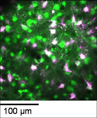
Visualizing Astrocyte Activation
A field of neurons (green) and astrocytes (purple) within the visual cortex, visualized by high-resolution two-photon imaging in the intact ferret brain. The astrocytes are labeled with the marker dye SR101, and both neurons and astrocytes are subsequently labeled with the calcium indicator OGB1. Activated astrocytes appear white. Image credit: James Schummers and Hongbo Yu, Laboratory of Mriganka Sur, MIT
As for the role of astrocytes in hemodynamics, Schummers and colleagues used optical imaging, under the same conditions as the two-photon measurements, to measure blood flow. They used the glutamate transporter antagonist DL-threo-β-benzyloxyaspartate (TBOA) to selectively silence astrocyte responses to visual stimuli while leaving neuronal responses intact. In the presence of TBOA, local blood flow responses were eliminated. “After decades of hemodynamic brain imaging, we are finally able to visualize the cellular mediator of vascular responses in action,” write Wolf and Kirchhoff.
This work most likely has far-reaching consequences beyond the visual cortex. Sur strongly believes that astrocytes are behaving in the same way in other areas of the brain, including the hippocampus (see, e.g., Diamond et al., 1998) and that this may be why fMRI and other hemodynamic imaging analysis works. “One direct implication of our work is that the specificity of brain imaging is made possible because of the specificity of astrocyte function,” he said.—Tom Fagan
References
Paper Citations
- Diamond JS, Bergles DE, Jahr CE. Glutamate release monitored with astrocyte transporter currents during LTP. Neuron. 1998 Aug;21(2):425-33. PubMed.
Further Reading
No Available Further Reading
Primary Papers
- Schummers J, Yu H, Sur M. Tuned responses of astrocytes and their influence on hemodynamic signals in the visual cortex. Science. 2008 Jun 20;320(5883):1638-43. PubMed.
- Wolf F, Kirchhoff F. Neuroscience. Imaging astrocyte activity. Science. 2008 Jun 20;320(5883):1597-9. PubMed.
Annotate
To make an annotation you must Login or Register.
Comments
The new findings of Schummers, Yu, and Sur are an important and interesting contribution to our understanding of the physiological role of astrocytes in brain function in vivo. Their findings provide strong evidence that rather than acting as a support network for neurons, astrocytes may be part of a neurovascular functional unit, playing an active role in information processing. By performing two-photon imaging of calcium signals in ferret visual cortex, they find that astrocytes display distinct spatial receptive fields and sharp tuning to visual features such as orientation and spatial frequency. (This finding is highly reminiscent of a prior study of Kelly and Van Essen [1974], who showed using microelectrode recording that glial cells in the primary visual cortex responded to visual stimuli with slow graded depolarizations, and that many of them showed a preference for a stimulus orientation similar to the optimal orientation for adjacent neurons.) Interestingly, the tuning of the astrocyte to these responses is even higher than that of the neurons, suggesting that they are important participants and controllers of the functional response. Moreover, when they pharmacologically block the activation of glutamate transporter current in astrocytes (that is normally stimulated by neuronal presynaptic glutamate release), they powerfully block the local vascular dilatation associated with visual sensory stimuli. (Similarly, Gurden et al. [2006] found in the olfactory bulb that presynaptic glutamate release and uptake by astrocytes form a critical pathway through which neural activity is linked to metabolic processing and hence to functional imaging signals, and that astrocyte glutamate transporters played a key role in activating an astrocyte intracellular calcium response.) Significantly, they show that specific visual stimuli induce focal astrocyte activation rather than glial calcium wave activity, and that neighboring astrocytes behave relatively independently of each other. Because activated astrocytes may release neuroactive substances (though probably not glutamate) that act on synapses or alter synaptic responsiveness, it will be important in the future to investigate the role of astrocyte activation on neuronal responses to visual stimuli.
The present findings strongly suggest that astrocytes play a critical role in information processing in the brain and that a cellular circuit of neurons, astrocytes, and blood vessels work together to control brain function. It is interesting that in a mouse model of Alzheimer disease, there is poor vasodilation in response to sensory stimuli (Takano et al., 2007). These investigators suggested that in this mouse model of Alzheimer disease, abnormal astrocytic activity may contribute to vascular instability in AD and thereby to neuronal demise. Given that reactive astrocytes have been found in neurodegenerative diseases (such as ALS) to strongly downregulate their glutamate transporters, the new findings of Schummers et al. help to explain the findings of Takano et al. (2007). Astrocytes lacking glutamate transporters would be predicted to be unable to elevate their intracellular calcium levels and thus unable to induce local vasodilation. If such an anomaly is more generalized throughout an Alzheimer’s affected brain (as would be predicted by the widespread gliosis), this could help to explain the widespread dysfunction of cognitive circuits in this disease.
References:
Kelly JP, Van Essen DC. Cell structure and function in the visual cortex of the cat. J Physiol. 1974 May;238(3):515-47. PubMed.
Gurden H, Uchida N, Mainen ZF. Sensory-evoked intrinsic optical signals in the olfactory bulb are coupled to glutamate release and uptake. Neuron. 2006 Oct 19;52(2):335-45. PubMed.
Takano T, Han X, Deane R, Zlokovic B, Nedergaard M. Two-photon imaging of astrocytic Ca2+ signaling and the microvasculature in experimental mice models of Alzheimer's disease. Ann N Y Acad Sci. 2007 Feb;1097:40-50. PubMed.
Make a Comment
To make a comment you must login or register.