Monomeric Seeds and Oligomeric Clouds—Proteopathy News from AAIC
Quick Links
Researchers studying protein aggregation have long debated two questions: What is the smallest structure than can seed different protein aggregates, aka strains, and how many different strains form within a brain? Researchers answered both at this year’s Alzheimer’s Association International Conference, held July 16-20 in London. Marc Diamond, University of Texas Southwestern Medical Center, Dallas, reported that single molecules of monomeric tau have the wherewithal to seed if certain motifs are exposed, while Mathias Jucker, University of Tübingen, Germany, presented evidence for “clouds” of different Aβ strains in the human brain. Other researchers thought the former offered a solid mechanistic explanation for seeding, and that the latter could have profound implications for amyloid imaging and therapy.

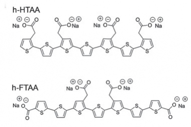
Fluorescent Probes.
Detection of different conformers of Aβ is possible with fluorescent dyes—conjugated oligothiophenes such as heptamer formyl thiophene acetic acid (h-FTAA) and heptamer hydrogen thiophene acetic acid. [Courtesy of Choong et al., Biofilms and Microbiomes 2016.]
Researchers in Jucker's lab spotted these clouds while using a type of dye to probe the structure of Aβ aggregates. Developed by Peter Nilsson and Per Hammarström at Linköping University, Sweden, luminescent conjugated oligothiophenes (LCOs) have a flexible backbone that conforms to protein structures that they bind. This molecular contortion changes the emission spectra of the LCOs. The dyes can theoretically identify dozens of β-sheet conformations because each will fluoresce differently. In effect, said Jucker, the spectrum is indicative of the amyloid structure.
Nilsson, Jucker, and colleagues have used these dyes to detect different forms of Aβ plaque in transgenic mouse models of AD and to distinguish them from fibrils of tau (see Aslund et al., 2009). In London, Jucker showed how two of these dyes detected a myriad of Aβ structures in human brain samples, as well.
Using a combination of two LCO dyes, Jay Rasmussen, Jasmin Mahler, and Nathalie Beschorner from the Jucker lab probed postmortem tissue from the frontal, temporal, and occipital cortices of 40 people who had had different types of AD. Twenty-one samples came from sporadic cases, three each from familial AD patients who carried APP V717I or PS A431E mutations, five from PS E280A FAD carriers, two from PS F105L patients, and six from patients with posterior cortical atrophy (PCA).

Spectral Plaque.
An LCO labels an amyloid plaque in a tissue section from an AD brain. Different colors represent different conformations of the plaque’s Aβ components. [Courtesy of Natalie Beschorner.]
While the mean spectral characteristics of plaques from different regions of a given brain looked similar, plaques from different mutation carriers had unique spectral features, which in turn differed from the spectra of plaques from sporadic AD and from PCA. Spectra of plaques from AD and PCA also differed. The heterogeneity did not stop there. When Jucker zoomed in on small areas within each plaque, many different individual spectra opened up before his eyes, indicative of many different conformations of Aβ. To quantify this, he measured the spectra from each of the two dyes at different regions in each plaque, using the ratio of the peak emission intensities as a measure of heterogeneity. He found dozens of different ratios across each plaque. “The data indicate that there are clouds of conformations of Aβ that are similar in regions of the same brain, but that differ between brains,” said Jucker.
Jucker does not yet know the exact structures to which these dyes bind. The spectra did not seem to correlate with the amount of Aβ in a plaque, or with whether the Aβ was resistant to proteases. “We need further analysis to explore the putative links between conformations detected by the dyes and phenotypes,” he said.
However, it does seem clear that these conformational clouds can be transmitted. The researchers injected extracts from PS A431E, APP V717I, and sporadic AD patients into APP transgenic mice. Months later LCOs detected clouds of different Aβ conformers in induced Aβ plaques in the brains of the mice, and these were similar overall to the LCO spectra of the source plaques in the human brain.
Researchers in the audience were intrigued. “This is really, really great work,” noted Diamond. He asked how it gibes with findings from Robert Tycko at the NIH, who reported that one type of Aβ structure predominantly forms when extracts from many regions of a single brain are used to seed amyloid fibrils in vitro (Sep 2013 news). “Does your result suggest that it may be very difficult to accurately amplify, in a single reaction, the diverse structures from the brain?” asked Diamond. Jucker agreed it might. “You could argue that Rob amplified the most important structure,” he suggested. Tycko was not at AAIC, but told Alzforum that his most recent data, which indicate heterogeneity among Aβ42 fibrils from various forms of AD, seems consistent with Jucker’s findings (Jan 2017 news).
Others wondered what Jucker’s finding means for amyloid PET scanning or for immunotherapy, particularly in a prevention paradigm where one might need an exquisitely specific antibody to prevent seeding and propagation of amyloid. Jucker said the heterogeneity could complicate both, further cautioning that the heterogeneity might be dynamic. “The seeds you see at end-stage disease may be very different from those you see early on,” he said.
Some of those concerns may not apply in cases where the seeds are monomers, and in the case of tau, that now seems more likely. Previously, researchers had concluded by extrapolating from the concentration of tau in solution that a monomer might be the smallest structure that could seed fibrillization (Chirita et al., 2005). In London, Diamond presented physical evidence that this is the case.
Diamond and colleagues have previously identified many different strains of tau in tissues samples from different tauopathies. These faithfully seed the propagation of identical strains in vitro and in vivo (May 2014 news). To determine the smallest seed that can pull off such a feat, Hilda Mirbaha in Diamond’s lab made tau fibrils in vitro, sonicated them, fractionated them by exclusion chromatography, and then tested the fragments in a cellular biosensor assay developed in Diamond’s lab (Oct 2014 news).
Mirbaha found that monomers derived from fibrillized tau seeded aggregation of tau in the cell assay. Curiously, monomers that had never been fibrillized could not. Mirbaha and co-workers ran extensive biophysical tests to ensure that these seeds were indeed monomers, including fluorescence correlation spectroscopy, which measures fluctuations in fluorescence intensity due to diffusion of molecules in solution to determine the size and number of particles present as they pass through an incident light beam.
Satisfied that monomers from fibrils made in vitro can act as seeds, the researchers then asked whether monomers from brain extracts do, as well. They captured tau aggregates from AD brain extracts using immunoprecipitation, gently homogenized them, and then separated tau species by exclusion chromatography. Again, fractions from AD brain containing only monomers seeded tau aggregation in cells. In contrast, monomer fractions from control brains did not. Interestingly, after sitting at room temperature for 24 hours, monomers from the AD but not control brain formed larger assemblies. “It appears we have two different types of monomer, one that likes to seed and self-assemble, and another inert monomer that does not,” said Diamond. The researchers have dubbed the recombinant seed competent and inert monomers Ms and Mi, respectively.
How do these monomers differ? Diamond and colleagues used cross-linking with mass spectrometry (XL-MS) to probe their structure. This technique identifies which parts of a structure lie adjacent to each other. The data indicated that Ms and Mi had distinct cross-linking patterns. In fact, one region around amino acids 150-275 seemed to cross-link solely in Ms. Based on the XL-MS data and the known structures of tau, Diamond worked with Lukasz Joachimiak, also at UT Southwestern, to model the structures of tau. These suggest that two motifs—VQIINK and VQIVYK, near the beginning of the second and third repeat domains, respectively—are accessible in Ms but buried in Mi.
Again, the audience was impressed. “I think the cross-link mass spec work is important, because it appears to identify the core element of the tau seed,” noted Benjamin Wolozin, Boston University. “VQIINK/VQIVYK were previously known to be important for tau aggregation. That in seed-competent tau these are solvent-accessible is very interesting, because it presents a concrete mechanism through which seeding becomes enabled,” Wolozin added (von Bergen at al., 2000; Apr 2017 news).
How do these monomer structures relate to strains? Diamond will investigate this. He believes the relative exposure of the VQIINK and VQIVYK motifs might be germane. For example, VQIVYK forms part of the core of the recently derived tau structure, whereas VQIINK does not (Jul 2017 news).
Are specific monomers responsible for different strains in different tauopathies? Here it gets complicated. Diamond has isolated monomers and larger seeds from AD and corticobasal dementia brain samples. All the seeds from the AD sample yielded one morphogenic strain in cells. Whole extracts from CBD brains yielded two strains, but Ms from CBD yielded those two strains plus a third. Diamond went on to show that monomers from CBD could apparently morph to give rise to at least three different strains. “A single monomer in CBD appears to account for the different strains we observe,” Diamond told Alzforum. “The critical idea is that there is a dominant superstructure of the monomer, but that it has some 'flexibility' to sample other shapes that, when assembled, form unique strains,” he added. By contrast, the “superstructure” of the AD monomer is relatively rigid, and give rise to just one strain.
Diamond believes there are hierarchies of tau conformation based on seeding competency. He showed a representative “family tree” of tau strains, with seed-competent strains branching into AD and CBD, and CBD strains branching further into at least three specific species. “This concept helps us pick apart how we can have the different types of tauopathy, and has implications for imaging, diagnosis, and treatment,” Diamond said.—Tom Fagan
References
News Citations
- Does Aβ Come In Strains? Glimpse Into Human Brain Suggests Yes
- Do Palettes of Aβ Fibril Strains Differ Among Alzheimer’s Subtypes?
- Like Prions, Tau Strains Are True to Form
- Cellular Biosensor Detects Tau Seeds Long Before They Sprout Pathology
- Does Tau’s Third Repeat Propagate Misfolding in Vivo?
- Tau Filaments from the Alzheimer’s Brain Revealed at Atomic Resolution
Paper Citations
- Aslund A, Sigurdson CJ, Klingstedt T, Grathwohl S, Bolmont T, Dickstein DL, Glimsdal E, Prokop S, Lindgren M, Konradsson P, Holtzman DM, Hof PR, Heppner FL, Gandy S, Jucker M, Aguzzi A, Hammarström P, Nilsson KP. Novel pentameric thiophene derivatives for in vitro and in vivo optical imaging of a plethora of protein aggregates in cerebral amyloidoses. ACS Chem Biol. 2009 Aug 21;4(8):673-84. PubMed.
- Chirita CN, Congdon EE, Yin H, Kuret J. Triggers of full-length tau aggregation: a role for partially folded intermediates. Biochemistry. 2005 Apr 19;44(15):5862-72. PubMed.
- von Bergen M, Friedhoff P, Biernat J, Heberle J, Mandelkow EM, Mandelkow E. Assembly of tau protein into Alzheimer paired helical filaments depends on a local sequence motif ((306)VQIVYK(311)) forming beta structure. Proc Natl Acad Sci U S A. 2000 May 9;97(10):5129-34. PubMed.
Other Citations
Further Reading
Papers
- Wegenast-Braun BM, Skodras A, Bayraktar G, Mahler J, Fritschi SK, Klingstedt T, Mason JJ, Hammarström P, Nilsson KP, Liebig C, Jucker M. Spectral discrimination of cerebral amyloid lesions after peripheral application of luminescent conjugated oligothiophenes. Am J Pathol. 2012 Dec;181(6):1953-60. PubMed.
Annotate
To make an annotation you must Login or Register.

Comments
No Available Comments
Make a Comment
To make a comment you must login or register.