Take It to the Brain Bank—Human Array Tomography Could Pay Dividends
Quick Links
Before cognition falters, Alzheimer’s disease wreaks havoc on synapses, the brain’s subcellular communication hubs. Yet synapses are tiny—too small to stain or count accurately using traditional light microscopes. Researchers have adapted array tomography, a high-resolution imaging method developed for rodent models (Micheva and Smith, 2007), to study the number, size, and protein composition of synapses in whole, postmortem human brain. Now, in the June 20 Nature Protocols, researchers led by Tara Spires-Jones at Massachusetts General Hospital, Boston, formally lay out the procedure, offering tips and troubleshooting advice in the hope that other labs will adopt the technology. They emphasize how specimens can be processed in parallel for array tomography (AT) and transmission electron microscopy (TEM). "Incorporation of AT and TEM into brain banking is a potent way of phenotyping synapses in well-characterized clinical cohorts in order to develop clinic-pathological correlations at the synapse level," they note.
In array tomography, whole tissues are embedded in resin and then sliced into ultrathin (70 nm) sections that can be imaged by immunofluorescence. The technique dramatically improves the axial resolution of light microscopy (1,000 nm), which is insufficient for visualizing individual synapses. Previously, the MGH researchers used AT to visualize Aβ oligomers at synaptic spines in mouse models of AD (ARF related conference story on Koffie et al., 2009) and to monitor mitochondria in tauopathy mice (Kopeikina et al., 2011). Other labs have used the technique to study synapses in songbirds (Oberti et al., 2011), zebrafish (Robles et al., 2011), and mice modeling epilepsy (Lacey et al., 2012).
More recently, Spires-Jones and colleagues adapted AT for studies in postmortem brain tissue from AD patients, showing that apolipoprotein E targets Aβ oligomers to synapses (Koffie et al., 2012; see also ARF SfN 2010 story). Another research group has used AT to look at the distribution of collagen in the human optic nerve (Winkler et al., 2010).
Preparing human brain samples for AT and TEM takes three days, but requires just two to three hours of extra labor time beyond typical brain banking procedures, Spires-Jones told Alzforum. Her lab is using AT to explore the role of tau at synapses. Though prior work suggests tau mediates Aβ-induced synapse loss, “nobody has been able to look at intact human brain to see if Aβ and tau are at the same synapse,” said Spires-Jones. The MGH researchers are also testing the role of glia in synaptic loss (see images below) and looking for synaptic α-synuclein in postmortem brain samples of people with dementia with Lewy bodies (DLB).
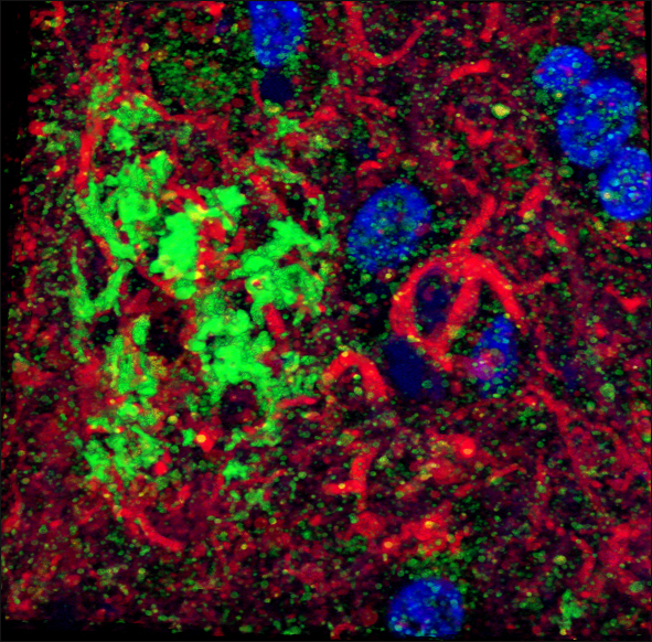
Three-dimensional reconstruction of superior temporal gyrus from an AD patient shows astrocytic processes (red) invading an Aβ plaque (green). DNA is stained with DAPI (blue). Image courtesy of Alberto Serrano-Pozo

Tripartite synapses are stained with presynaptic marker VGLUT1 (blue), postsynaptic marker PSD 95 (green), and astrocytic end foot marker EAAT1 (red). Image courtesy of Alberto Serrano-Pozo
In 2011, Stephen Smith and Kristina Micheva—who pioneered the AT method at Stanford University School of Medicine, Palo Alto, California—founded the Silicon Valley company Aratome LLC to process samples for AT. About 20 labs worldwide are using AT today, Smith told Alzforum. Though most studies are still in mice, there is increasing interest in human AT, he said. Spires-Jones is collaborating with MGH colleagues, as well as researchers in the U.K. and Spain, on rodent and human AT studies.—Esther Landhuis
References
News Citations
- Spine Shrinkers: Aβ Oligomers Caught in the Act
- San Diego: Aβ Oligomers Seen, With ApoE, at Synapses of Human Brain
Paper Citations
- Micheva KD, Smith SJ. Array tomography: a new tool for imaging the molecular architecture and ultrastructure of neural circuits. Neuron. 2007 Jul 5;55(1):25-36. PubMed.
- Koffie RM, Meyer-Luehmann M, Hashimoto T, Adams KW, Mielke ML, Garcia-Alloza M, Micheva KD, Smith SJ, Kim ML, Lee VM, Hyman BT, Spires-Jones TL. Oligomeric amyloid beta associates with postsynaptic densities and correlates with excitatory synapse loss near senile plaques. Proc Natl Acad Sci U S A. 2009 Mar 10;106(10):4012-7. PubMed.
- Kopeikina KJ, Carlson GA, Pitstick R, Ludvigson AE, Peters A, Luebke JI, Koffie RM, Frosch MP, Hyman BT, Spires-Jones TL. Tau accumulation causes mitochondrial distribution deficits in neurons in a mouse model of tauopathy and in human Alzheimer's disease brain. Am J Pathol. 2011 Oct;179(4):2071-82. PubMed.
- Oberti D, Kirschmann MA, Hahnloser RH. Projection neuron circuits resolved using correlative array tomography. Front Neurosci. 2011;5:50. PubMed.
- Robles E, Smith SJ, Baier H. Characterization of genetically targeted neuron types in the zebrafish optic tectum. Front Neural Circuits. 2011;5:1. PubMed.
- Lacey CJ, Bryant A, Brill J, Huguenard JR. Enhanced NMDA receptor-dependent thalamic excitation and network oscillations in stargazer mice. J Neurosci. 2012 Aug 8;32(32):11067-81. PubMed.
- Koffie RM, Hashimoto T, Tai HC, Kay KR, Serrano-Pozo A, Joyner D, Hou S, Kopeikina KJ, Frosch MP, Lee VM, Holtzman DM, Hyman BT, Spires-Jones TL. Apolipoprotein E4 effects in Alzheimer's disease are mediated by synaptotoxic oligomeric amyloid-β. Brain. 2012 Jul;135(Pt 7):2155-68. PubMed.
- Winkler M, Jester B, Nien-Shy C, Massei S, Minckler DS, Jester JV, Brown DJ. High resolution three-dimensional reconstruction of the collagenous matrix of the human optic nerve head. Brain Res Bull. 2010 Feb 15;81(2-3):339-48. PubMed.
Other Citations
External Citations
Further Reading
Papers
- Koffie RM, Hashimoto T, Tai HC, Kay KR, Serrano-Pozo A, Joyner D, Hou S, Kopeikina KJ, Frosch MP, Lee VM, Holtzman DM, Hyman BT, Spires-Jones TL. Apolipoprotein E4 effects in Alzheimer's disease are mediated by synaptotoxic oligomeric amyloid-β. Brain. 2012 Jul;135(Pt 7):2155-68. PubMed.
- Kopeikina KJ, Carlson GA, Pitstick R, Ludvigson AE, Peters A, Luebke JI, Koffie RM, Frosch MP, Hyman BT, Spires-Jones TL. Tau accumulation causes mitochondrial distribution deficits in neurons in a mouse model of tauopathy and in human Alzheimer's disease brain. Am J Pathol. 2011 Oct;179(4):2071-82. PubMed.
- Koffie RM, Meyer-Luehmann M, Hashimoto T, Adams KW, Mielke ML, Garcia-Alloza M, Micheva KD, Smith SJ, Kim ML, Lee VM, Hyman BT, Spires-Jones TL. Oligomeric amyloid beta associates with postsynaptic densities and correlates with excitatory synapse loss near senile plaques. Proc Natl Acad Sci U S A. 2009 Mar 10;106(10):4012-7. PubMed.
Primary Papers
- Kay KR, Smith C, Wright AK, Serrano-Pozo A, Pooler AM, Koffie R, Bastin ME, Bak TH, Abrahams S, Kopeikina KJ, McGuone D, Frosch MP, Gillingwater TH, Hyman BT, Spires-Jones TL. Studying synapses in human brain with array tomography and electron microscopy. Nat Protoc. 2013 Jul;8(7):1366-80. PubMed.
Annotate
To make an annotation you must Login or Register.
Comments
No Available Comments
Make a Comment
To make a comment you must login or register.