Synaptic Metropolis—Microscopic Census Documents 300,000 Protein Inhabitants
Quick Links
Wielding a combination of imaging and quantification techniques, researchers have generated the most detailed three-dimensional map of the synapse to date, showing how approximately 300,000 proteins squeeze into this tiny space. As reported in the May 30 Science, most of the proteins are involved in synaptic vesicle trafficking. Included in the mix are thousands of copies of amyloid precursor protein (APP), which blanket the extracellular surface of the synaptosome (see video below). There is also an abundance of α-synuclein on the cytosol side. The investigators hope the model will serve as a baseline that allows researchers to pinpoint synapse problems in neurodegenerative diseases, said senior author Silvio Rizzoli from the University of Göttingen in Germany.

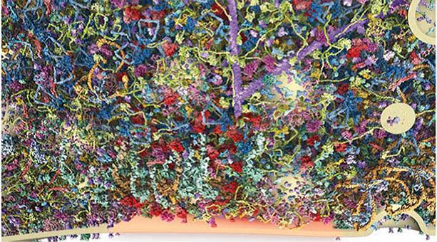
The Fabric of a Synapse. A section of a synaptic bouton, containing 60 different proteins, shows crisscrossing actin filaments (purple, from top), and vesicles (cream, right) that travel toward the synapse (bottom, orange). Courtesy of Wilhelm et al., 2014, Science.
Rizzoli likened the synaptosome to a jungle. “If you want to know how a jungle functions, you are not going to stop at finding out what species exist there,” he said. “Unless you know how they interact, you can’t figure out how it operates.” His other reason for creating the model was simpler. “I just wanted to see what it would look like,” he said.
First author Benjamin Wilhelm and colleagues started by fractionating cellular extracts from the cortex and cerebellum of a rat brain, enriching for synaptosomes. Then they quantified the concentrations of 62 known synaptic proteins by immunoblot. The researchers used mass spectrometry to quantify an additional 1,100 proteins within the preparation. To determine how many copies of each protein were in the synaptosome, the researchers first counted the number of synaptosomes in their preparation using electron and fluorescence microscopy. Then, with the concentration and mass of each protein in hand, the researchers calculated the copy number of each protein per synaptosome.
Rizzoli said the most striking result from this initial accounting was the sheer abundance of proteins known to be involved in the fusion of synaptic vesicles to the cell membrane, or exocytosis. For example, a single synaptosome contained upward of 20,000 copies of each of the SNARE proteins involved in exocytosis, even though only one to three copies of each SNARE are needed for one vesicle fusion event, and no more than five vesicles are thought to fuse at once, Rizzoli said. In contrast, proteins involved in recycling vesicles, or endocytosis, were far less abundant. Rizzoli hypothesized that synaptosomes contain an overabundance of exocytic proteins simply because neurons must be able to fire on demand. “You have to have an enormous excess of the proteins that can do it,” he said.
With the copy number of various proteins in hand, the researchers next wanted to see how they were distributed throughout the synaptosome. To do this, they used stimulated emission depletion (STED), a high-resolution fluorescence microscopy technique that allows visualization down to about 40 nm resolution, roughly the size of a synaptic vesicle (see Apr 2006 news story). They used STED to measure the distribution of each protein in the synaptosome, then mapped out the positions of 60 different proteins, which together made up some 300,000 protein copies. Wilhelm integrated protein structures, as well as information from the literature about the way certain proteins interact with each other, to create a virtual three-dimensional map. It includes the synaptosome plasma membrane, synaptic vesicles, and the cytosol (see video, best viewed in full-screen mode).
In the video, the fuzzy purple protein coating the synaptosome surface is none other than APP, which the researchers report exists at 6,000 copies per synaptosome. This protein stands out more than others on the surface because it is the only one with a large extracellular domain, Rizzoli said. He was surprised to find so many copies of APP there, but declined to speculate about the reason for APP’s ample representation on the synaptosome surface. Only about 100 copies of BACE were identified. Inside the synaptosome, α-synuclein—represented by nearly 4,000 copies—was found mingling with vesicles.
Rizzoli hopes that researchers studying neurodegenerative diseases will use this synaptosome as a baseline model to evaluate the integrity of synaptosomes from animal models of disease. Researchers could analyze diseased synaptosomes by mass spectrometry, Rizzoli said, and compare their protein complement to the baseline. Rizzoli said he talked with the Michael J. Fox Foundation about trying this with a rat model of Parkinson’s disease.—Jessica Shugart
References
News Citations
Further Reading
Papers
- Zhang WI, Röhse H, Rizzoli SO, Opazo F. Fluorescent in situ hybridization of synaptic proteins imaged with super-resolution STED microscopy. Microsc Res Tech. 2014 Jul;77(7):517-27. Epub 2014 Apr 10 PubMed.
- Denker A, Bethani I, Kröhnert K, Körber C, Horstmann H, Wilhelm BG, Barysch SV, Kuner T, Neher E, Rizzoli SO. A small pool of vesicles maintains synaptic activity in vivo. Proc Natl Acad Sci U S A. 2011 Oct 11;108(41):17177-82. Epub 2011 Sep 8 PubMed.
Primary Papers
- Wilhelm BG, Mandad S, Truckenbrodt S, Kröhnert K, Schäfer C, Rammner B, Koo SJ, Claßen GA, Krauss M, Haucke V, Urlaub H, Rizzoli SO. Composition of isolated synaptic boutons reveals the amounts of vesicle trafficking proteins. Science. 2014 May 30;344(6187):1023-8. PubMed.
Annotate
To make an annotation you must Login or Register.

Comments
Washington University
Clearly, this is an amazing paper. The detail of the number and spatial location of proteins is remarkable. I appreciate their discussion of what the copy number of proteins may mean functionally; to compensate for a relatively small amount of endocytic machinery, the presynaptic terminal contains a vast excess of prepared synaptic vesicles.
Applying this approach to all kinds of systems now, including AD, should yield incredible information. My bias will be to see how much APP is present and where it is located within the terminal.…More
View all comments by John CirritoMake a Comment
To make a comment you must login or register.