First Primate Model of Familial AD: Marmosets with Presenilin Mutation
Quick Links
Mice fail to model many aspects of Alzheimer’s disease. Could monkeys capture the disease more fully? In a preprint posted to bioRxiv on August 24, researchers led by Takaomi Saido, RIKEN Center for Brain Science, Wako, and Erika Sasaki, Central Institute for Experimental Animals, Kawasaki, both in Japan, report the first nonhuman primate genetic model of AD. The authors used gene editing to delete exon 9 in the presenilin 1 gene of marmosets, a small South American monkey that has more similarities to people in their brain structure and physiology than rodents do. This deletion occurs in some people with familial AD.
Three marmosets with one copy of the edited PS1 gene were born. Fibroblasts from the young animals make more Aβ42 than do cells from wild-type controls, though it remains to be seen if the marmosets will develop plaques and tangles as they age.
Other researchers were enthusiastic. “This is a major advance in generating an animal model that more accurately reflects human disease,” Cynthia Lemere at Brigham and Women’s Hospital, Boston, wrote to Alzforum (full comment below). Lary Walker at Emory University, Atlanta, agreed. “These PSEN1-ΔE9 marmosets are an exciting new model. If they prove to develop a truly AD-like phenotype, they will be a significant addition to the armamentarium of researchers who are fighting to eradicate the disease,” he wrote (full comment below).

Better AD Model? The common marmoset lives 10 to 15 years; its sleep patterns and some social behaviors resemble those of people. [© Raimond Spekking / CC BY-SA 4.0 (via Wikimedia Commons).]
Mouse models of amyloidosis do not develop neurofibrillary tangles or neurodegeneration, nor can they reflect most of the cognitive and behavioral changes that afflict people with AD. By contrast, the common marmoset, Callithrix jacchus, has complex social behaviors and sleep patterns that resemble ours. Their metabolism and immune systems are also more similar to those in people than those in rodents. Moreover, marmosets can live 10 to 15 years in captivity, and typically develop hyperphosphorylated tau and amyloid plaques as they age. Their Aβ sequence is identical to the human protein. Finally, raising and maintaining research colonies is feasible because these primates are small and produce many offspring. They typically weigh less than a pound.
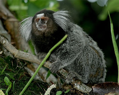
Aging Problems. The common marmoset can develop hyperphosphorylated tau and amyloid plaques in the brain as it ages. [Courtesy of Georges Néron, Wikimedia.]
To edit the marmoset genome, joint first authors Kenya Sato and Hiroki Sasaguri used transcription activator-like effector nucleases (TALEN) rather than CRISPR, because the former method works more efficiently and uniformly with marmosets, producing less genetic mosaicism (Hockemeyer et al., 2011; Sato et al., 2016). Because homozygous presenilin mutations were embryonic lethal, as they are in mice, the authors targeted haploid marmoset ova before fertilization. Deletion of the splice acceptor site at the 3' end of exon 9 produced presenilin lacking this exon. The authors then fertilized 578 of these genetically altered ova with wild-type marmoset sperm, and implanted 154 surviving zygotes into 77 surrogate mothers. Six babies survived till birth; three of those carried the presenilin 1 mutation.
The three young marmosets, two male and one female, are all heterozygous for the mutation, but carry no obvious off-target genetic alterations. The authors analyzed fibroblasts taken from earlobe tissue, and found the cells pumped out more Aβ42 than did wild-type fibroblasts, more than doubling their Aβ42/40 ratio. This is similar to human fibroblasts taken from PS1 mutation carriers (Lemere et al., 1996; Scheuner et al., 1996; Kumar-Singh et al., 2006).
If amyloidosis progresses similarly as it does in people with the mutation, the marmosets might be expected to develop amyloid plaques after two to three years, rather than the seven years it takes in wild-types, the authors noted.
They speculate that marmosets may be more likely than mouse models to develop neurofibrillary tangles, because their longer lifespans give more time for these deposits to form. However, Lemere noted that wild-type vervets, an Old World monkey, develop abundant plaques but few tangles even over a lifespan of 20 to 30 years. “It is possible that other factors, such as immune responses to toxins, infections, etc., contribute to those changes. This new marmoset model will facilitate answering these kinds of mechanistic questions, as well as allow for drug screening,” she wrote.
The authors plan to make this marmoset model widely available to the research community.—Madolyn Bowman Rogers
References
Paper Citations
- Hockemeyer D, Wang H, Kiani S, Lai CS, Gao Q, Cassady JP, Cost GJ, Zhang L, Santiago Y, Miller JC, Zeitler B, Cherone JM, Meng X, Hinkley SJ, Rebar EJ, Gregory PD, Urnov FD, Jaenisch R. Genetic engineering of human pluripotent cells using TALE nucleases. Nat Biotechnol. 2011 Aug;29(8):731-4. PubMed.
- Sato K, Oiwa R, Kumita W, Henry R, Sakuma T, Ito R, Nozu R, Inoue T, Katano I, Sato K, Okahara N, Okahara J, Shimizu Y, Yamamoto M, Hanazawa K, Kawakami T, Kametani Y, Suzuki R, Takahashi T, Weinstein EJ, Yamamoto T, Sakakibara Y, Habu S, Hata J, Okano H, Sasaki E. Generation of a Nonhuman Primate Model of Severe Combined Immunodeficiency Using Highly Efficient Genome Editing. Cell Stem Cell. 2016 Jul 7;19(1):127-38. Epub 2016 Jun 30 PubMed.
- Lemere CA, Lopera F, Kosik KS, Lendon CL, Ossa J, Saido TC, Yamaguchi H, Ruiz A, Martinez A, Madrigal L, Hincapie L, Arango JC, Anthony DC, Koo EH, Goate AM, Selkoe DJ, Arango JC. The E280A presenilin 1 Alzheimer mutation produces increased A beta 42 deposition and severe cerebellar pathology. Nat Med. 1996 Oct;2(10):1146-50. PubMed.
- Scheuner D, Eckman C, Jensen M, Song X, Citron M, Suzuki N, Bird TD, Hardy J, Hutton M, Kukull W, Larson E, Levy-Lahad E, Viitanen M, Peskind E, Poorkaj P, Schellenberg G, Tanzi R, Wasco W, Lannfelt L, Selkoe D, Younkin S. Secreted amyloid beta-protein similar to that in the senile plaques of Alzheimer's disease is increased in vivo by the presenilin 1 and 2 and APP mutations linked to familial Alzheimer's disease. Nat Med. 1996 Aug;2(8):864-70. PubMed.
- Kumar-Singh S, Theuns J, Van Broeck B, Pirici D, Vennekens K, Corsmit E, Cruts M, Dermaut B, Wang R, Van Broeckhoven C. Mean age-of-onset of familial alzheimer disease caused by presenilin mutations correlates with both increased Abeta42 and decreased Abeta40. Hum Mutat. 2006 Jul;27(7):686-95. PubMed.
External Citations
Further Reading
Primary Papers
- Sato K, Sasaguri H, Kumita W, Inoue T, Kurotaki Y, Nagata K, Mihira N, Sato K, Sakuma T, Yamamoto T, Tagami M, Manabe R, Ozaki K, Okazaki Y, Saido TC, Sasaki E. A non-human primate model of familial Alzheimer’s disease. bioRxiv, August 24, 2020
Annotate
To make an annotation you must Login or Register.
Comments
Ann Romney Center for Neurologic Diseases, Brigham and Women's Hospital and Harvard Medical School
This is a big achievement. I realize it is early days but the data reported by Sato et al. in bioRxiv on August 24, indicate that RIKEN Center for Brain Science, in collaboration with the Central Institute for Experimental Animals, has generated the first-ever nonhuman primate (NHP) model of familial AD using Transcription Activator-Like Effector Nuclease (TALEN) gene editing to remove the 3' splice site of exon 9 of the presenilin 1 gene (PSEN1) in New World marmosets. As of April 2019, the investigators have generated three monoallelic PSEN1-DE9 marmosets and three wild-type PSEN1 siblings. Biallelic deletion of exon 9 led to embryonic lethality, similar to that seen in mice. Off-target effects and mosaicism were absent in the PSEN1-DE9 marmosets. PSEN1 N- and C-terminal fragments were reduced by about half in the mutant marmosets, which still carry one wild-type PSEN1 allele. Conditioned media from earlobe-derived fibroblasts demonstrated a twofold increase in the Aβ42/40 ratio.…More
Whether the monoallelic deletion of PSEN1 exon 9 will be transmitted as a germline mutation remains to be determined as the animals are not yet sexually mature. However, based on another TALEN-induced marmoset model, it looks likely that this PSEN1-DE9 genotype will be carried forward with successive breeding. This is a major advance in generating an animal model that more accurately reflects human disease due to its strong homology with human genes, similar sleep patterns, visual and auditory learning and memory, and long life span (10-15 years). The authors predict the PSEN1-DE9 marmosets will have accelerated Aβ deposition starting around 2-3 years of age instead of about 7 years in wild-type animals.
Although marmosets develop hyperphosphorylated tau in their brains with aging, they, like other NHPs, do not tend to develop neurofibrillary tangles (NFTs). Whether accelerating Aβ deposition will induce NFTs remains to be determined. Our work (Lemere et al., 2004) and that of Latimer et al. (Latimer et al., 2019) indicate that Old World Caribbean vervets, which live 20-30 years, develop abundant cerebral Aβ plaques and less-abundant phosphorylated tau inclusions, but only rarely develop NFTs, even in the presence of high numbers of plaques and a long life span. It is possible that other factors, such as immune responses to toxins, infections, etc., contribute to those changes. This new marmoset model will facilitate answering these kinds of mechanistic questions as well as allow for drug screening. Kudos to Drs. Sato, Saido, and Sasaki for this major step forward in Alzheimer’s research!
References:
Lemere CA, Beierschmitt A, Iglesias M, Spooner ET, Bloom JK, Leverone JF, Zheng JB, Seabrook TJ, Louard D, Li D, Selkoe DJ, Palmour RM, Ervin FR. Alzheimer's disease abeta vaccine reduces central nervous system abeta levels in a non-human primate, the Caribbean vervet. Am J Pathol. 2004 Jul;165(1):283-97. PubMed.
Latimer CS, Shively CA, Keene CD, Jorgensen MJ, Andrews RN, Register TC, Montine TJ, Wilson AM, Neth BJ, Mintz A, Maldjian JA, Whitlow CT, Kaplan JR, Craft S. A nonhuman primate model of early Alzheimer's disease pathologic change: Implications for disease pathogenesis. Alzheimers Dement. 2019 Jan;15(1):93-105. Epub 2018 Nov 19 PubMed.
Emory University
Few scientists in the Alzheimer's field would disagree that we need an animal model that more faithfully manifests the disease as it occurs in humans. Genetically modified mice have been a boon to studies of cellular and molecular mechanisms, but they have been poor predictors of the efficacy of treatments. A number of animal species naturally develop Aβ plaques in old age, and some develop neurofibrillary tangles, but full-blown, human-like Alzheimer's disease—including dementia and widespread plaques and tangles in the brain—has not yet been found in any nonhuman organism.
Thus, this report by Sato and colleagues of the initial development of a genetically modified nonhuman primate model of hereditary Alzheimer's disease is of considerable interest. The animals are common marmosets (Callithrix jacchus), a New World monkey that is native to South America. Although they are evolutionarily more distant from humans than are commonly studied Old World primates (such as rhesus monkeys), they have a number of advantages as potential models of AD: 1) They naturally express human-sequence Aβ, and they develop Aβ plaques, cerebral Aβ-amyloid angiopathy, and a degree of hyperphosphorylated tauopathy (but not bona fide neurofibrillary tangles) as they age; 2) they are small (roughly the weight of rats), but they have a large brain:body weight ratio and a relatively sophisticated behavioral repertoire; and 3) compared to most other primates, marmosets reproduce prolifically, and their maximum lifespan is comparatively short (~16 years). …More
Using transcription activator-like effector nuclease (TALEN), Sato et al. deleted exon 9 of the gene for presenilin-1 (PSEN1-ΔE9; a mutation that is associated with a hereditary form of early onset AD and a selective increase in Aβ42). Notably, the animals are otherwise normal genetically. Hence, the altered PSEN1 acts by influencing the APP/Aβ that is innately generated by the marmosets (rather than transgenic human APP, which is required in rodent models to yield human-sequence Aβ). In this sense, the genetic makeup of the monkeys fairly closely resembles that in humans bearing the PSEN1-ΔE9 mutation.
The early characterization of biomarkers in the young marmosets is promising. The expression of altered PSEN1 is nicely demonstrated, and no off-target effects of the genetic manipulation were detected. As in humans with the PSEN1-ΔE9 mutation, the Aβ42:40 ratio (assessed in marmoset fibroblasts) is elevated. The researchers plan a comprehensive phenotypic analysis as the animals age, including behavior, imaging, and biomarkers. Importantly, they make the welcome proposal to provide PSEN1-ΔE9 marmosets to other researchers once this becomes feasible.
Ultimately, the value of the PSEN1-ΔE9 marmoset model will hinge on the degree to which the behavioral and pathologic changes mimic those in Alzheimer's disease. If AD-like plaques and tangles do occur in the brain, the time course of pathogenesis still must be factored into the practicality of marmosets as models. A favorable scenario might be the appearance of plaques by 2-3 years of age (as suggested by the authors), and then tangles and cognitive decline by age 4 or 5. Even with this ambitious timeline, the expense of relatively long-term studies in sufficiently large numbers of marmosets could exceed the resources of many laboratories. In addition, the breeding and care of marmosets requires specialized infrastructure and expertise that few research facilities have available.
That said, better animal models are undoubtedly needed to accelerate research on basic mechanisms, and to reliably test the efficacy of new treatment and diagnostic strategies. In late-stage preclinical testing of therapeutic agents, the marmosets might serve the dual purpose of assessing efficacy as well as safety in a non-rodent species. These PSEN1-ΔE9 marmosets are an exciting new model. If they prove to develop a truly AD-like phenotype, they will be a significant addition to the armamentarium of researchers who are fighting to eradicate the disease.
Indiana University School of Medicine
Development of an unprecedented nonhuman primate model (NHP) of familial Alzheimer’s disease (FAD) by Saido and colleagues is an outstanding accomplishment. As others mentioned on Alzforum, a familial NHP model is a significant contribution to the AD field and urgently needed to develop interventions for this disease. It is becoming more apparent that rodent models, including humanized transgenic AD mice and rats, although valuable for our basic understanding of AD, are falling short as the key to discover effective drugs that would be successful in human clinical trials and demonstrate wide treatment efficacy in the broader patient population.
Indeed, a marmoset model that is available to the research community would facilitate the global fight against AD by providing an animal model with a biological background much closer to that found in humans than can be accomplished by using rodents. Use of primates, in general, is not new to the field of neurodegeneration and provides unique opportunities for translational research in AD (Haque and Levey, 2019). A matched set of relationships among neuropathological, cerebrospinal fluid, imaging, and behavioral modalities consistent with early AD, support future use of the vervet monkey to discover disease mechanisms, biomarkers, and novel therapeutic strategies (Latimer et al., 2019). …More
However, we should approach this field with cautious optimism as NHPs do not get AD. Nevertheless, non-transgenic NHPs have contributed to the understanding of the role of nongenetic factors, such as environmental toxins and epigenetic factors. For example, the role of depleted norepinephrine has recently been tested in adult and aged rhesus macaques to develop a potential model for testing AD interventions (Duffy et al., 2019).
Notably, non-transgenic NHPs have provided important clues to how early life exposure contributes to late-life disorders in general (Wu et al., 2008). Such research formed the basis for the Latent Early-life Associated Regulation (LEARn) model to offer an epigenetic explanation for idiopathic neurobiological diseases (Lahiri et al., 2009). Work in etiology, behavior, and molecular mechanisms mimicking AD conditions would be challenging to replicate in rodents (Maloney and Lahiri, 2016). In other words, familial PS1 or not, NHPs deserve increased attention and resources to advance the field. Of course, this must be balanced against the much higher economic, regulatory, and ethical costs involved in the use of NHP models. However, those could be looked at as a strong incentive for efficient and meaningful experimental designs. Taken together, both nongenetic and familial NHP models would be a significant leap for AD therapeutics.
References:
Haque RU, Levey AI. Alzheimer's disease: A clinical perspective and future nonhuman primate research opportunities. Proc Natl Acad Sci U S A. 2019 Dec 23; PubMed.
Latimer CS, Shively CA, Keene CD, Jorgensen MJ, Andrews RN, Register TC, Montine TJ, Wilson AM, Neth BJ, Mintz A, Maldjian JA, Whitlow CT, Kaplan JR, Craft S. A nonhuman primate model of early Alzheimer's disease pathologic change: Implications for disease pathogenesis. Alzheimers Dement. 2019 Jan;15(1):93-105. Epub 2018 Nov 19 PubMed.
Duffy KB, Ray B, Lahiri DK, Tilmont EM, Tinkler GP, Herbert RL, Greig NH, Ingram DK, Ottinger MA, Mattison JA. Effects of Reducing Norepinephrine Levels via DSP4 Treatment on Amyloid-β Pathology in Female Rhesus Macaques (Macaca Mulatta). J Alzheimers Dis. 2019;68(1):115-126. PubMed.
Wu J, Basha MR, Brock B, Cox DP, Cardozo-Pelaez F, McPherson CA, Harry J, Rice DC, Maloney B, Chen D, Lahiri DK, Zawia NH. Alzheimer's disease (AD)-like pathology in aged monkeys after infantile exposure to environmental metal lead (Pb): evidence for a developmental origin and environmental link for AD. J Neurosci. 2008 Jan 2;28(1):3-9. PubMed.
Lahiri DK, Maloney B, Zawia NH. The LEARn model: an epigenetic explanation for idiopathic neurobiological diseases. Mol Psychiatry. 2009 Nov;14(11):992-1003. PubMed.
Maloney B, Lahiri DK. Epigenetics of dementia: understanding the disease as a transformation rather than a state. Lancet Neurol. 2016 Jun;15(7):760-74. Epub 2016 May 9 PubMed.
Make a Comment
To make a comment you must login or register.