Up Close and Exciting: The NMDA Glutamate Receptor
Quick Links
One of several types of ligand-gated ion channel, the N-methyl-D-aspartate (NMDA) receptor transmits excitatory stimuli in the brain. When calcium flows through this channel, neural plasticity happens and learning and memory are facilitated. Alas, too much of this good thing leads to excitotoxicity that might cause neurodegeneration in diseases such as Alzheimer’s, Parkinson’s, and Huntington’s. While widely accepted, all this is complex and visually murky. A paper in the May 30 Science affords the most detailed view of the intact NMDA receptor yet. Erkan Karakas and Hiro Furukawa of Cold Spring Harbor Laboratory, New York, used X-ray crystallography to peer at the arrangement of the receptor’s four subunits and their various domains. Their data confirm the previously predicted configuration of its subunits and suggest an explanation for why allosteric modulators have an impact on the regions furthest from the ion channel itself. Unravelling the minute details of this receptor will help researchers understand its complex function and how it might be better targeted by therapies, wrote the authors. Memantine, an NMDA receptor antagonist, is one of only four drugs approved for AD.
“The NMDA receptor is so important for disease, and there has been an enormous effort to design different compounds that bind to its various regions,” Furukawa told Alzforum. “This full-length structure will help us understand which parts of the NMDA receptors these compounds are binding.”

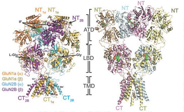
NMDAR Details: The overall structure of the NMDA receptor (left) shows how the GluN1 and N2 subunits form its amino terminal domain (ATD), ligand binding domain (LBD), and transmembrane domain (TMD). Compare to the previously resolved AMPA receptor (right). Image courtesy of Science/AAAS.
Previous studies have visualized only bits and pieces of the receptor, because it has been difficult to express it in its entirety and in large enough quantities to perform crystallography. Karakas and Furukawa overcame these difficulties with genetic tweaks. They then crystallized NMDA receptors that were bound to two different agonists—glycine or L-glutamate—or the allosteric inhibitor ifenprodil. The scientists stabilized the heterotetramer by creating disulfide bonds between the four subunits. X-ray crystallography then revealed the atomic structure of the protein and interactions between the receptor’s four subunits, including both extracellular and transmembrane portions, at a resolution of 4 angstroms.
It turns out that the NMDA receptor looks a bit like a hot-air balloon. The extracellular amino terminal domain (ATD) and ligand-binding domain (LBD) form the balloon portion and the transmembrane domain makes up the basket. As previously predicted, the four subunits—two identical pairs—lie in a staggered arrangement akin to two white and two black squares that make a bigger square on a checkerboard. This is likely because of steric clashes in the ligand-binding domain that would occur if pairs butted up against each other, the authors wrote.
The scientists compared their new conquest to the AMPA receptor, whose structure had been solved previously (Sobolevsky et al., 2009). They saw that the close proximity of the NMDAR’s ATD, which binds allosteric modulators, and LBD, which controls channel opening, explains why allosteric modulators influence when the channel opens. In essence, structural changes from the ATD can be easily transmitted to the LBD. In the AMPA receptor, the ATD and LBD interact less to confer more of a Y-shape, and the ATD does not serve as a site for allosteric modulation.
Furukawa said that his team will solve the structures for different states of the ion channel when NMDA binds to various agonists, antagonists, and allosteric modulators. This should help researchers improve therapeutic compounds, including ifenprodil and memantine, which bind the ATD and channel pore, respectively. (Unlike memantine, ifenprodil is not a therapeutic.) It may even help in the design of new compounds, Furukawa told Alzforum.—Gwyneth Dickey Zakaib
References
Therapeutics Citations
Paper Citations
- Sobolevsky AI, Rosconi MP, Gouaux E. X-ray structure, symmetry and mechanism of an AMPA-subtype glutamate receptor. Nature. 2009 Dec 10;462(7274):745-56. PubMed.
Further Reading
Papers
- Kvist T, Steffensen TB, Greenwood JR, Mehrzad Tabrizi F, Hansen KB, Gajhede M, Pickering DS, Traynelis SF, Kastrup JS, Bräuner-Osborne H. Crystal structure and pharmacological characterization of a novel N-methyl-D-aspartate (NMDA) receptor antagonist at the GluN1 glycine binding site. J Biol Chem. 2013 Nov 15;288(46):33124-35. Epub 2013 Sep 26 PubMed.
- Yao Y, Belcher J, Berger AJ, Mayer ML, Lau AY. Conformational analysis of NMDA receptor GluN1, GluN2, and GluN3 ligand-binding domains reveals subtype-specific characteristics. Structure. 2013 Oct 8;21(10):1788-99. Epub 2013 Aug 22 PubMed.
- Kumar J, Mayer ML. Functional insights from glutamate receptor ion channel structures. Annu Rev Physiol. 2013;75:313-37. Epub 2012 Sep 4 PubMed.
Primary Papers
- Karakas E, Furukawa H. Crystal structure of a heterotetrameric NMDA receptor ion channel. Science. 2014 May 30;344(6187):992-7. PubMed.
Annotate
To make an annotation you must Login or Register.

Comments
NIMH
Even though the atomic structure of the NMDA receptor-channel complex in parts has been described or modelled in detail and the molecular correlates dictating its functionality are generally understood, this elegant study by Karakas and Furukawa presents a major further development. It confidently lines up with the work by Hayashi, Watkins, Nowak, Sobolevsky, and co-workers on this intriguing and highly important protein (channel) and is likely to have major implications for the field of synaptic biology and several specialized areas of neuroscience research. It is reassuring that the now fully deciphered three-dimensional structure of the NMDA heterotetramer reveals no major surprises but confirms and consolidates the knowledge already gained over several decades of research.
Make a Comment
To make a comment you must login or register.