Artificial Human Blood Vessels: A Model for Cerebral Amyloid Angiopathy?
Quick Links
Can blood vessels grown from human cells shed light on amyloid buildup in the Alzheimer’s brain? In the October 10 eLife, researchers led by Cheryl Wellington at the University of British Columbia, Vancouver, Canada, argue they can. They seeded a tube-shaped scaffold with human endothelial cells, smooth muscle cells, and astrocytes to create artificial arteries through which they pumped media. To model cerebral amyloid angiopathy, an accumulation of Aβ in brain blood vessel walls, they added Aβ40 and Aβ42. Both peptides accumulated in the vessel tissue, eventually forming fibrils. But the cells also transported some of the Aβ into the vessel lumen, with ApoE and high-density lipoprotein preferentially facilitating transport of Aβ42 over Aβ40.
- Scientists engineered artificial arteries from human endothelial cells, smooth muscle cells, and astrocytes.
- Aβ42 and Aβ40 accumulate in, move across, the vessel walls.
- ApoE and HDL preferentially facilitate clearance of Aβ42 over Aβ40.
Some researchers criticized this model, but others welcomed it. “It is a step forward. In vitro studies have mostly relied on cells grown on a flat surface without any of the biomechanical characteristics related to blood flow,” said Costantino Iadecola, Weill Cornell Medical College in New York. “That Aβ42 was transported more easily than Aβ40 is novel,” he added.

Lab Artery. Side (left) and luminal (right) views of a 2 mm inner diameter artificial artery. [Courtesy of Cheryl Wellington.]
Scientists have been engineering artificial blood vessels for decades. While some simply co-cultured endothelial and smooth muscle cells in a dish, others created three-dimensional tubes that mimicked vessel anatomy and tolerated fluid pressures encountered in the body. These more realistic models range from capillary networks grown on microchannels etched into stiff or pliable matrices to hollow fibers seeded with endothelial cells through which media courses in bursts to simulate pulsing blood flow (e.g., Huang and Niklason, 2014; Tsvirkun et al., 2017; Lamberti et al., 2014). A commercial kit available through Flocel Inc., Cleveland, includes endothelial cells and astrocytes that model the blood-brain barrier (Cucullo et al., 2007). With these tissue engineering advances and the availability of patient-derived stem cells to model vascular aspects of specific diseases, research with artificial vessels is growing (e.g., Atchison et al., 2017; Dash et al., 2016; Truskey and Fernandez, 2015).

Pump It Up.
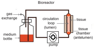
Media (orange) flows in pulsating waves through an artificial human blood vessel (pink) suspended in medium (tan). Researchers add molecules or cells to the luminal or anteluminal compartments. [Courtesy of Robert et al., eLife.]
To study cerebral amyloid angiopathy (CAA), first author Jerome Robert upgraded a model he had designed to study atherosclerosis. He added astrocytes to an artificial vessel that comprised only endothelial and smooth muscle cells (Robert et al., 2013). Robert seeded the lumen of a tubular, biodegradable polymer scaffold with human umbilical cord myofibroblasts, i.e. smooth muscle cell precursors. He maintained the scaffold in a bioreactor chamber in which the scaffold’s lumen formed part of a circulation loop, separate from the fluid bathing the outer surface, or anteluminal side (see image above). After allowing the fibroblasts to adhere for three days, Robert turned on the circulation for a week. Then he added human umbilical cord endothelial cells to the luminal side. After giving the cells a few days to settle, he switched on the pump again and ramped up the flow rate over the next two weeks. “The flow helps create a mature tissue,” he explained. It encourages cells to secrete extracellular matrix components, make tight endothelial connections, and properly polarize. The approach resembles one that generates vessel-like tubes to repair malformed vessels in people (Schmidt et al., 2006). It yields layers of smooth muscle cells surrounding an endothelial lining impermeable to macromolecules.

Artificial Vessel. Cross-section of an artificial human vessel (left) made of endothelial cells (yellow) and smooth muscle cells (tan). To make it resemble brain blood vessels (right), researchers added astrocytes (blue). [Courtesy of Robert et al., eLife.]
To make the vessels more like those in the brain, Robert added primary human astrocytes to the anteluminal side of the vessel, after the endothelial cells had settled in. The astrocytes encircled the smooth muscle layer. Somehow sensing the presence of the astrocytes, the endothelial cells began expressing blood-brain barrier proteins, including tight junction proteins and the Glut-1 glucose transporter. The researchers also saw astrocytes putting out protrusions similar to the endfeet that encircle endothelial cells in the brain.
“The approach is a classic one, seeding cells onto a scaffold. The major novel aspect here is that they included multiple cell types,” said Mohammad Kiani of Temple University, Philadelphia, who has built artificial microvascular networks. Wellington said that to her knowledge, this is the first fully human cerebral vessel engineered to model CAA. “With a 2 mm internal diameter and layers of smooth muscle cells, rather than the pericytes associated with small vessels, these artificial vessels resemble the brain’s leptomeningeal and penetrating arteries, which are particularly affected in CAA,” she said.
The field needs such models, Wellington argues, because mice cannot fully recapitulate the properties of human blood vessels. For example, in mouse blood, high-density lipoproteins (HDL) outnumber low-density lipoproteins (LDL), whereas the opposite is true in people. And whereas mice have one APOE allele, people have three. “A powerful aspect of this technology is its ability to incorporate human tissues,” wrote Courtney Lane-Donovan and Joachim Herz, University of California, San Francisco, in an accompanying commentary.
The CAA field has experienced its share of challenges studying disease mechanisms directly in patients. Guojun Bu at the Mayo Clinic in Jacksonville, Florida, noted that, though helpful, observations of postmortem brain tissue or, in a few cases, tracking markers in accessible tissues such as cerebrospinal fluid, have yielded only limited, correlative data.
To model amyloid accumulation, the researchers added Aβ40 and Aβ42 into the anteluminal chamber. As per ELISA and immunostaining, the peptides lodged in the vessel walls and began forming fibrils within hours. The vessels allowed only half the amount of peptides to cross into the lumen that a cell-free scaffold did.
Suspecting lipoproteins might affect this, the researchers turned their attention to ApoE and high-density lipoprotein (HDL). A previous study from Berizlav Zlokovic’s lab at the University of Southern California in Los Angeles reported that ApoE, particularly ApoE4, blunts Aβ clearance across the mouse BBB (Deane et al., 2008), while Wellington had reported that HDL reduced AD risk and dialed down soluble brain Aβ levels in the APPPS1 mouse model of AD (Robert et al, 2016).
Welllington’s group tested the effect of ApoE in both models. They added recombinant ApoE to the antelumen of artificial vessels comprising only endothelial and smooth muscle cells. To test the effects of endogenous ApoE, they added GW3965, a drug that increases the expression of the apolipoprotein, to vessels wrapped in astrocytes, the major producers of ApoE in the brain. In the bipartite system, ApoE2 boosted Aβ clearance and levels of Aβ42 in the vessel tissue fell.
Wellington noted that this contrasted with Zlokovic’s findings. She offered various reasons, including differences between mice and people and between whole-brain vasculature and specific vessels. Like ApoE, HDL also lowered Aβ42 in the vessels and, most strikingly, HDL and ApoE together had a synergistic effect. Integrating results from multiple combinations of vessel types and treatments, the authors concluded that the HDL and ApoE combo helped both Aβ42 and Aβ40 cross over into the lumen, with peptide levels rising there as they dropped in the vessel wall.
Interestingly, Wellington found that ApoE2 and HDL smoothed the transit of Aβ42 more than of Aβ40, consistent with the known predominance of Aβ40 over Aβ42 in blood vessels in familial cases of CAA (Alonzo et al., 1998). Bu noted Aβ40 may diffuse through the brain parenchyma more easily than does Aβ42, and thus find its way to blood vessels more readily. The issue remains unresolved, he said.
Robert and colleagues found that while ApoE2 facilitated amyloid transport, ApoE4 did not. However, paired with HDL from healthy volunteers, both ApoE isoforms, as well as ApoE3 produced by astrocytes in the tripartite model, boosted clearance. The results may vary with HDL from other sources, since HDL can be compromised by aging and cardiovascular disease (Riwanto and Landmesser, 2013). Wellington thinks it will take time to sort out HDL’s contributions given its multiple roles in vessel health and disease.
While intrigued by the study, researchers also pointed out limitations. Iadecola and Damir Janigro, founder of Flocel Inc., lamented the model’s lack of a perivascular space. “This is not the way it is in the human brain,” said Janigro. The astrocytes in the artificial vessel contact smooth muscle cells directly, without the basement membrane proteins and macrophages usually found in the space between these two cell types. Iadecola said the shortcoming diminishes the artificial vessel’s value as a model for CAA because the perivascular space likely serves as an important route for Aβ elimination (Carare et al., 2008; Bakker et al, 2016).
Wellington agreed that engineering a perivascular space is an important next step. “We are consulting with others to help us evaluate potential solutions, including biomaterial, bioprinting, and microfluidic approaches,” she wrote to Alzforum.
Others noted that the artificial vessel cannot recapitulate glymphatic flow or perivascular drainage (May 2016 news). “It is difficult to see how the model could be used to test intramural, periarterial drainage of Aβ along the basement membranes of capillaries and arteries,” wrote Roxana Carare and Roy Weller at the University of Southampton, England. One problem is that the endothelial LRP receptors that have been implicated in the transport of Aβ42 across the BBB are present in capillary walls, not arteries. “While the endothelial cells used in this model have features of a BBB, their umbilical cord origin and the absence of the other cerebral cells such as pericytes most likely result in an extracellular matrix that is not specific to the cerebral vasculature,” they added.
Could Robert’s technique be used to engineer capillaries or smaller vessels, given their relevance to Aβ removal? Kiani doubts it, because it is challenging to make such tiny scaffolds and rig them up for circulation. Microfluidics techniques, which allow researchers to build networks of channels tens to hundreds of micrometers in diameter, would probably work better, he said.
Kiani suggested using endothelial cells from the human aorta, which is more similar to brain arteries than umbilical cord veins. His group had trouble reproducing the behavior of brain microvascular endothelial cells using endothelial cells derived from cord veins, but he emphasized his system was different from Wellington’s. Bu would like to see patient-derived astrocytes used to produce ApoE2 or ApoE4.
Is the model perfect? “No,” said Iadecola, “but you have to start somewhere. The vasculature is too complicated to model everything at once, and they have succeeded with the basics.” Bu and Janigro complimented the use of human cells and pulsatile flow, and Bu said being able to independently manipulate the luminal and anteluminal sides of a vessel is valuable. Researchers were also excited about the potential for carrying out long-term studies. “They cultured the vessels for several weeks; that is pretty impressive,” said Kiani. Given this longevity, Bu believes the model might be able to reproduce pathology and replicate the subsequent degeneration of smooth muscle cells in CAA.
Iadecola wondered how the artificial vessel might stack up to excised vessel fragments as models of disease. “Ex vivo preparations are very useful, but you can’t control many factors,” said Wellington. For example, the use of induced pluripotent stem cells from patients in building an artificial vessel provides many ways of experimentally tweaking the system, she said. Wellington expects artificial vessels to yield more reproducible results than excised vessels. This would be critical for testing candidate therapies, as she hopes to do.
Looking ahead, Wellington hopes to add components such as neurons and microglia to the model. “We are taking the first steps in a long process toward better experimental platforms,” she said.
“One goal is to help train others. We want to have people visit the lab. It’s not something you can learn in one weekend, so we want to create ways to enable others to use the model and have it validated,” she said.—Marina Chicurel
References
Research Models Citations
News Citations
Paper Citations
- Huang AH, Niklason LE. Engineering of arteries in vitro. Cell Mol Life Sci. 2014 Jun;71(11):2103-18. Epub 2014 Jan 8 PubMed.
- Tsvirkun D, Grichine A, Duperray A, Misbah C, Bureau L. Microvasculature on a chip: study of the Endothelial Surface Layer and the flow structure of Red Blood Cells. Sci Rep. 2017 Mar 24;7:45036. PubMed.
- Lamberti G, Prabhakarpandian B, Garson C, Smith A, Pant K, Wang B, Kiani MF. Bioinspired microfluidic assay for in vitro modeling of leukocyte-endothelium interactions. Anal Chem. 2014 Aug 19;86(16):8344-51. Epub 2014 Aug 5 PubMed.
- Cucullo L, Hossain M, Rapp E, Manders T, Marchi N, Janigro D. Development of a humanized in vitro blood-brain barrier model to screen for brain penetration of antiepileptic drugs. Epilepsia. 2007 Mar;48(3):505-16. Epub 2007 Feb 22 PubMed.
- Atchison L, Zhang H, Cao K, Truskey GA. A Tissue Engineered Blood Vessel Model of Hutchinson-Gilford Progeria Syndrome Using Human iPSC-derived Smooth Muscle Cells. Sci Rep. 2017 Aug 15;7(1):8168. PubMed.
- Dash BC, Levi K, Schwan J, Luo J, Bartulos O, Wu H, Qiu C, Yi T, Ren Y, Campbell S, Rolle MW, Qyang Y. Tissue-Engineered Vascular Rings from Human iPSC-Derived Smooth Muscle Cells. Stem Cell Reports. 2016 Jul 12;7(1):19-28. PubMed.
- Truskey GA, Fernandez CE. Tissue-engineered blood vessels as promising tools for testing drug toxicity. Expert Opin Drug Metab Toxicol. 2015 Jul;11(7):1021-4. Epub 2015 May 31 PubMed.
- Robert J, Weber B, Frese L, Emmert MY, Schmidt D, von Eckardstein A, Rohrer L, Hoerstrup SP. A three-dimensional engineered artery model for in vitro atherosclerosis research. PLoS One. 2013;8(11):e79821. Epub 2013 Nov 14 PubMed.
- Schmidt D, Asmis LM, Odermatt B, Kelm J, Breymann C, Gössi M, Genoni M, Zund G, Hoerstrup SP. Engineered living blood vessels: functional endothelia generated from human umbilical cord-derived progenitors. Ann Thorac Surg. 2006 Oct;82(4):1465-71; discussion 1471. PubMed.
- Deane R, Sagare A, Hamm K, Parisi M, Lane S, Finn MB, Holtzman DM, Zlokovic BV. apoE isoform-specific disruption of amyloid beta peptide clearance from mouse brain. J Clin Invest. 2008 Nov 13; PubMed.
- Robert J, Stukas S, Button E, Cheng WH, Lee M, Fan J, Wilkinson A, Kulic I, Wright SD, Wellington CL. Reconstituted high-density lipoproteins acutely reduce soluble brain Aβ levels in symptomatic APP/PS1 mice. Biochim Biophys Acta. 2016 May;1862(5):1027-36. Epub 2015 Oct 9 PubMed.
- Alonzo NC, Hyman BT, Rebeck GW, Greenberg SM. Progression of cerebral amyloid angiopathy: accumulation of amyloid-beta40 in affected vessels. J Neuropathol Exp Neurol. 1998 Apr;57(4):353-9. PubMed.
- Riwanto M, Landmesser U. High density lipoproteins and endothelial functions: mechanistic insights and alterations in cardiovascular disease. J Lipid Res. 2013 Dec;54(12):3227-43. Epub 2013 Jul 20 PubMed.
- Carare RO, Bernardes-Silva M, Newman TA, Page AM, Nicoll JA, Perry VH, Weller RO. Solutes, but not cells, drain from the brain parenchyma along basement membranes of capillaries and arteries: significance for cerebral amyloid angiopathy and neuroimmunology. Neuropathol Appl Neurobiol. 2008 Apr;34(2):131-44. Epub 2008 Jan 16 PubMed.
- Bakker EN, Bacskai BJ, Arbel-Ornath M, Aldea R, Bedussi B, Morris AW, Weller RO, Carare RO. Lymphatic Clearance of the Brain: Perivascular, Paravascular and Significance for Neurodegenerative Diseases. Cell Mol Neurobiol. 2016 Mar;36(2):181-94. Epub 2016 Mar 18 PubMed.
Further Reading
Papers
- Bang S, Lee SR, Ko J, Son K, Tahk D, Ahn J, Im C, Jeon NL. A Low Permeability Microfluidic Blood-Brain Barrier Platform with Direct Contact between Perfusable Vascular Network and Astrocytes. Sci Rep. 2017 Aug 14;7(1):8083. PubMed.
- Castellano JM, Kim J, Stewart FR, Jiang H, DeMattos RB, Patterson BW, Fagan AM, Morris JC, Mawuenyega KG, Cruchaga C, Goate AM, Bales KR, Paul SM, Bateman RJ, Holtzman DM. Human apoE isoforms differentially regulate brain amyloid-β peptide clearance. Sci Transl Med. 2011 Jun 29;3(89):89ra57. PubMed.
Primary Papers
- Robert J, Button EB, Yuen B, Gilmour M, Kang K, Bahrabadi A, Stukas S, Zhao W, Kulic I, Wellington CL. Clearance of beta-amyloid is facilitated by apolipoprotein E and circulating high-density lipoproteins in bioengineered human vessels. Elife. 2017 Oct 10;6 PubMed.
- Lane-Donovan C, Herz J. Building a better blood-brain barrier. Elife. 2017 Oct 10;6 PubMed.
Annotate
To make an annotation you must Login or Register.

Comments
University of Southampton School of Medicine
University of Southampton School of Medicine
There is firm evidence from experimental studies and observations on human brains with Alzheimer’s disease that cerebral amyloid angiopathy is a consequence of a failure of clearance of Aβ and interstitial fluid from the brain. In order to test new therapeutic strategies, there is an urgent need for new in vitro models that test the clearance of Aβ. The present study represents a significant step toward efficient in vitro models, as it mimics the walls of large cerebral arteries and the transport of Aβ radially across the wall of the blood vessel into the blood. From an anatomical point of view, the astrocytes are not present in leptomeningeal arteries, but rather leptomeningeal sheets cover the vessels. It is difficult to see how the model could be used to test intramural periarterial drainage of Aβ along the basement membranes of capillaries and arteries. One problem is that transport into the blood occurs at the capillary level, as the LRP transporters are present in the capillary wall and there is no evidence of transport of Aβ across the walls of arteries into the blood. While the endothelial cells used in this model have features of a blood-brain barrier, their umbilical cord origin and the absence of the other cerebral cells such as pericytes most likely result in an extracellular matrix that is not specific to the cerebral vasculature.
Make a Comment
To make a comment you must login or register.