Calcium Sensor STIM2 Maintains Synapses, Ebbs in Alzheimer’s
Quick Links
In Alzheimer’s disease, cognitive decline correlates most closely with the loss of synapses, but exactly what makes them vanish remains a mystery. Now, in the April 2 Neuron, researchers led by Ilya Bezprozvanny at the University of Texas Southwestern Medical Center, Dallas, tie synapse loss to perturbed calcium regulation in dendritic spines. The scientists found that neurons from mice that express an AD gene make less stromal interaction molecule 2. STIM2 is a calcium sensor. When it is lacking, less calcium enters spines, and mature synapses are not stabilized. Intriguingly, neurons from old mice also have less STIM2, as do samples from human brains with sporadic Alzheimer’s disease. “We think this may be the common mechanism for spine loss in aging and AD,” Bezprozvanny told Alzforum.
“I’m very excited about this paper. It represents a new contribution to the field by analyzing synapse loss from a mechanistic perspective,” said Grace (Beth) Stutzmann at Rosalind Franklin University in North Chicago, Illinois. She was not involved in the work, but noted that the data could lead to fresh therapeutic approaches. “Preserving synapses makes sense as a therapeutic strategy, because it gets at the heart of what’s driving the cognitive loss,” she said.
Numerous studies have shown that calcium regulation goes awry in Alzheimer’s disease, triggered by either presenilin mutations or amyloid accumulation (see Jul 2008 news story; Jul 2008 news story; Oct 2008 webinar). Too much calcium can have toxic effects on neurons, including spine loss (see Feb 2010 news story; Nov 2010 news story; Jul 2011 news story).
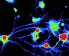
Calcium influx in hippocampal neurons. [Image courtesy of Sun et al., Neuron 2014, Fig 2a.]
To further investigate how calcium induces spine loss, Bezprozvanny and colleagues used knock-in mice that express the human presenilin 1 mutation PS1-M146V. Previously, Bezprozvanny had reported that familial AD mutations such as this one disrupted presenilins’ ability to act as calcium leak channels in the endoplasmic reticulum (ER) (see Sep 2006 news story), which unbalances calcium regulation in neurons (see Jun 2010 news story). The idea of a presenilin ER calcium leak function has been controversial but was recently independently validated (see Bandara et al., 2013). The PS1-M146V knock-ins do not express human amyloid precursor protein or accumulate human Aβ42. Since mouse Aβ is not toxic to synapses, this allowed the researchers to focus on the effects of calcium.
First author Suya Sun found that by six months of age, knock-in mice had only half the number of mushroom spines on hippocampal neurons as did controls. Scientists believe that mushroom spines represent mature synapses that store memories (see Bourne and Harris, 2007). These little knobs are lost in AD and to a lesser extent in normal aging (see Dickstein et al., 2013). In live imaging experiments, mushroom spines formed normally in the knock-in neurons but were later lost, suggesting the cell cannot stabilize these structures, Bezprozvanny told Alzforum.
The authors traced the problem to a lack of STIM2. STIM2 sits in the membrane of ER sections that run adjacent to the cell membrane. There, its internal portion senses calcium levels in the ER and its cytosolic portion makes contact with calcium channels on the cell membrane. When ER calcium levels fall, STIM2 triggers these channels to open and allow more calcium into the cell.
The authors found that STIM2 occupies mushroom spines, but not immature “thin” spines. Hippocampal neurons of 6-month-old knock-in mice had half as many STIM2-positive mushroom spines as did controls. The authors saw a similar loss of hippocampal STIM2 in 9-month-old APPPS1 mice, demonstrating that the findings were not unique to the knock-in model. Wild-type mice lost hippocampal STIM2 starting at 12 months old, suggesting that a similar process occurs during normal aging.
To look more closely at the consequences of this, the authors developed a conditional STIM2 knockout mouse that lacked the protein only in some hippocampal neurons. As expected, these neurons lost mushroom spines. Spines lacking STIM2 brought in 85 percent less calcium than did normal spines—a severe impairment in calcium regulation.
What does low calcium do to a spine? Calcium stimulates CaMKII phosphorylation, thus activating it, and activated CaMKII turns on small GTPase proteins that reorganize the actin cytoskeleton to remodel spines. In both STIM2 knockout and PS1 knock-in neurons, the authors saw a drop in phosphorylated CaMKII and a failure to activate the GTPases Cdc42 and Rac1. The results suggest a role for this pathway in maintaining mature spines, Bezprozvanny said.
As expected, expressing a STIM2 transgene in PS1 knock-in neurons normalized calcium entry as well as CaMKII and GTPase activation, and that restored the number of mushroom spines to wild-type levels. Because the knock-in mice have only mild memory impairments, the authors were not able to look at behavioral changes. They plan to do this experiment in another mouse model, Bezprozvanny said.
To test if these results relate to sporadic AD, where there are no presenilin mutations, the authors examined postmortem brains from 11 AD patients and 11 age-matched controls. In cortical lysates from AD brains, STIM2 levels were down by half, and this correlated with lower scores on the Mini-Mental State Examination shortly before they died. In contrast, no drop in STIM2 was seen in six brains from people with mild cognitive impairment (MCI), suggesting that this loss occurs later in disease when memory is more impaired.
Assuming these data are confirmed in larger samples and independent studies, the findings beg the question of what makes STIM2 fall in AD and normal aging. Clearly this does not require a presenilin mutation. Bezprozvanny hypothesized that the cell dials down STIM2 expression in response to an abundance of calcium in the ER. Several different pathways affecting calcium may lead to this point, among them presenilin mutations or amyloid, Bezprozvanny said. In ongoing work, he is examining how this pathway is affected in mouse models that accumulate Aβ.
Commentators praised the thoroughness of the study. “The data are convincing and the effects robust,” said Kim Green at the University of California, Irvine. Green previously found that STIM2 levels dropped with age in human brains, agreeing with Bezprozvanny’s mouse results. Green found it intriguing that the PS1 knock-in mice lost even more STIM2 than old mice did. “That tells me that age-related changes in calcium could contribute toward the formation of the disease, but once you have the disease, that may further affect calcium pathways,” he said. It would be interesting to see if other proteins in these pathways fall with age or in Alzheimer’s, Green noted.
It is unclear how this mechanism might be targeted therapeutically. Several calcium channel blockers are used for heart disease, but because calcium is involved in so many cellular processes, attempts to regulate it might lead to undesirable side effects. “We might need more targeted, specific ways of modulating calcium activity in the brain,” said Brian Bacskai at Massachusetts General Hospital, Boston. One approach might be to find ways to turn up STIM2 expression, since this protein appears specific for dendritic spines. Another possibility is to develop selective activators of store-operated calcium influx channels in the mushroom spines, an approach that Bezprozvanny is pursuing. Researchers agreed that the new findings will pique interest in studying calcium biology in AD. “It brings calcium dysregulation as a unifying mechanism of AD to the forefront,” Bacskai said.—Madolyn Bowman Rogers
References
News Citations
- Pump It Up—Presenilins Linked to ER SERCA Activity
- More Calcium News: Plaques Cause Dendrite Damage via Ion Overload
- Calcium Hypothesis—Studies Beef Up NFAT, CaN, Astrocyte Connections
- Can Calcium Channel Blockers Save Stressed-Out Dopaminergic Neurons?
- New Clues to Calcium Toxicity in Familial AD
- Presenilins Open Escape Hatch for ER Calcium
- Perplexing Presenilins: New Evidence for Calcium Leak Channels
Webinar Citations
Research Models Citations
Paper Citations
- Bandara S, Malmersjö S, Meyer T. Regulators of calcium homeostasis identified by inference of kinetic model parameters from live single cells perturbed by siRNA. Sci Signal. 2013 Jul 9;6(283):ra56. PubMed.
- Bourne J, Harris KM. Do thin spines learn to be mushroom spines that remember?. Curr Opin Neurobiol. 2007 Jun;17(3):381-6. Epub 2007 May 10 PubMed.
- Dickstein DL, Weaver CM, Luebke JI, Hof PR. Dendritic spine changes associated with normal aging. Neuroscience. 2012 Oct 13; PubMed.
Further Reading
News
Primary Papers
- Sun S, Zhang H, Liu J, Popugaeva E, Xu NJ, Feske S, White CL 3rd, Bezprozvanny I. Reduced synaptic STIM2 expression and impaired store-operated calcium entry cause destabilization of mature spines in mutant presenilin mice. Neuron. 2014 Apr 2;82(1):79-93. PubMed.
Annotate
To make an annotation you must Login or Register.
Comments
No Available Comments
Make a Comment
To make a comment you must login or register.