AMYPAD Confirms: Amyloid PET Improves Diagnosis
Quick Links
The first reports from AMYPAD, a large European study of amyloid PET scans, agree with previous studies: The technology strongly affects Alzheimer’s diagnosis. At the Alzheimer’s Association International Conference, held July 31 to August 4 virtually and in San Diego, California, AMYPAD leaders said the scans changed doctors’ diagnosis of one in three people who came to memory clinics with cognitive complaints. However, during a six-month follow-up period, the scans made no difference in the patients’ quality of life, nor reduced their use of medical services, raising questions about the cost-effectiveness of this expensive imaging tool.
- Amyloid scans changed one-third of diagnoses in AMYPAD study.
- More brain amyloid correlated with faster cognitive decline.
- Adding tau imaging improved prognostic power.
AMYPAD data also confirmed that a positive scan, or even subthreshold amyloid accumulation, predicts future cognitive decline. This decline occurred mostly in a person’s memory, with little impact on executive or visuospatial function. Other talks at AAIC noted that the scans’ predictive power can be sharpened by adding tau PET and MRI. In small studies, the majority of cognitively normal participants with positive amyloid and tau scans and hippocampal shrinkage on MRI developed mild cognitive impairment over the next two to three years, compared with about 10 percent of those who were positive for amyloid only. This strengthens the case for using biomarkers of amyloid, tau, and neurodegeneration in the clinic to determine prognosis, speakers said.
Better Diagnosis, But Not Better Health
Several previous studies have assessed how amyloid PET affects clinical care. In the U.S., IDEAS reported that scans changed treatment plans in two-thirds of its 11,409 participants (Aug 2017 conference news; Nov 2018 conference news; Apr 2019 news). Smaller European studies found a lesser benefit, with about one-quarter of diagnoses changed (Nov 2016 news; Jun 2018 news). Even so, the clinical usefulness of amyloid scanning has not been fully established. Additional studies, such as IDEAS 2 focusing on underrepresented minorities in the U.S., are ongoing.
In San Diego, Daniele Altomare of the University of Geneva offered a first look at AMYPAD data. The project consists of a diagnostic and patient management study (DPMS) and a prognostic and natural history study (PNHS). DPMS investigated the clinical use of amyloid scans (Frisoni et al., 2019). It recruited 840 people who sought an evaluation at one of eight participating memory clinics in France, Germany, the Netherlands, Spain, Sweden, Switzerland, and the U.K. between April 2018 and October 2020. Nearly all were white.
In this cohort, 244 people had subjective cognitive decline, 341 MCI, and 255 dementia. More cognitively impaired patients tended to be older, with average ages of 69, 72, and 75 years, respectively, for the three groups. The SCD group had somewhat more years of education on average than the other groups, while the dementia group was more depressed than the others. These findings are typical for memory clinic patients (Altomare et al., 2022).
Participants were randomized to one of three arms. In the first, they underwent PET scanning right away as part of their diagnostic workup, and had a follow-up scan after 12 to 18 months. In the second, they were scanned after eight months. In the third, physicians chose whether and when to use amyloid scanning; on average, this group underwent scanning six weeks after coming to the clinic. For the main outcome measure, researchers compared physicians’ initial diagnostic impression with the diagnosis they had settled on three months later.
In arm 1, diagnosis changed in 44 percent of cases. In arm 3, where PET scans were done slightly later, diagnosis changed in 29 percent of cases, not statistically different from arm 1. This contrasts with arm 2, which had no PET scans at this point; there, diagnosis changed in 11 percent of cases. Thus, about a third more of the cohort had an altered diagnosis when PET scans were part of their workup. Diagnostic changes were almost always consistent with the PET results, Altomare said. He noted that physicians altered their assessment more often after a negative than a positive scan, suggesting the biomarker is particularly useful for ruling out AD.
Physicians became more confident in their diagnosis once they had a scan in hand. In arm 1, physicians expressed high confidence in 40 percent of their diagnoses after three months. In arm 3, this was 37 percent; in arm 2, 11 percent.
Alas, a presumably more precise diagnosis did not translate to any effects on health outcomes over six months. Patients in each arm rated their own quality of life similarly, and beyond the cost of the PET scans themselves, the three arms were the same in their use of healthcare resources. This implies that PET scans are not cost-effective over the short term, Altomare acknowledged. In answer to an audience question, he suggested that prescreening with blood biomarkers could help manage costs. He speculated that longer-term follow-up might yet reveal more health differences. The study will continue to track participants for at least a year after their baseline visit.
What is the psychological impact of disclosing amyloid PET scan results to people? The scientists found that positive scans caused some distress, with participants reporting they had more intrusive thoughts and hyperarousal afterward, and avoided thinking about the results. These symptoms did not reach the threshold for clinical concern, and were lower than those reported after learning of other serious health conditions, such as cancer, Altomare said. The findings reinforce other studies suggesting that people can handle learning their amyloid status (see related conference story).
Predicting Progression
Separately, AMYPAD examined the prognostic prowess of amyloid PET (Lopes Alves et al., 2020). The PNHS part of AMYPAD enrolled more than 1,500 participants, most of whom were at the preclinical or early prodromal stage of AD. In San Diego, David Vállez García of Amsterdam UMC reported preliminary findings from the first 1,044 participants. These came from six research cohorts: EPAD, the ALFA+ study at Barcelonaβeta Brain Research Center, FACEHBI in Spain, EMIF Twin 60+ and EMIF 90+ in the Netherlands, and DELCODE in Germany.
Upon amyloid scanning, 650 of these participants proved to be amyloid-negative, with centiloid values around zero. Another 267 were in a “gray zone,” with an average of 10 centiloids of plaque. The remaining 127 were amyloid-positive, with varying amounts of amyloid plaque that averaged more than 50 centiloids. As might be expected, the amyloid-positive group was older, more cognitively impaired, and more likely to carry an APOE4 allele than the others.
Tracking participants’ cognition for up to six years, the scientists found that the amount of amyloid a person had at baseline predicted his or her global cognitive decline on the MMSE, as well as decline on tests of immediate recall, recognition, and visual memory. It also predicted functional decline on the iADL, as well as on one measure of attention, but had no impact on executive or visuospatial function over this time frame. On the MMSE, immediate recall memory, and attention tests, decline got steeper over time.
As expected, demographic factors such as age, sex, and education played into how fast a person declined. Education was protective, age made decline worse, and sex had complex, mixed effects. Having an APOE4 allele hastened slippage on the MMSE. Vállez García plans to further analyze how these risk factors interact with plaque burden to influence prognosis. All AMYPAD data will be made available to the research community via the ADDI FAIR platform.
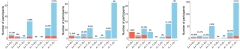
Predictive Power. In four different cohorts—PREVENT-AD, HABS, AIBL, and Knight ADRC (left to right)—people who were A+T+N+ were most likely to progress to MCI (red), while the majority of people who were only A+ remained cognitively healthy (blue). [Courtesy of Cherie Strikwerda-Brown.]
The PNHS findings dovetail with other studies shown at AAIC. Cherie Strikwerda-Brown of McGill University, Montreal, presented findings from four longitudinal studies of people who were cognitively healthy at baseline. The full cohort included 251 people seen at the Knight ADRC in St. Louis, 153 in the Harvard Aging Brain Study, 128 from PREVENT-AD in Montreal, and 48 from the Australian Imaging, Biomarker, and Lifestyle (AIBL) study. At baseline, participants underwent amyloid and tau PET and volumetric MRI as the measures for the current ATN classification of preclinical AD. Because each of the four studies used somewhat differently methodology, Strikwerda-Brown, working with Sylvia Villeneuve at McGill, analyzed each separately.
Over up to three years of follow-up, people who had all three markers—amyloid, tau, neurodegeneration—were the most likely to develop mild cognitive impairment, while those with none were the least. In PREVENT-AD, 57 percent of A+T+N+ individuals developed MCI; in HABS, 71 percent; in AIBL, everyone did. In the Knight ADRC, which used a CDR score of 0.5 or more to measure disease progression, 43 percent worsened. By contrast, people who were positive for amyloid, but not tau or brain atrophy, tended to stay stable, with fewer than one in 10 going on to MCI. On sensitive cognitive measures such as the RBANS and the PACC5 cognitive composite, A+T+ participants diverged from the others, declining much faster. Strikwerda-Brown concluded from this that the ATN criteria have prognostic value.
Likewise, Karly Cody of the University of Wisconsin-Madison reported that amyloid and tau both affect cognitive decline. UW researchers led by Sterling Johnson previously developed an amyloid “clock” to predict the rate of cognitive decline based on when a person became amyloid-positive (Oct 2019 news). To find out if adding tau PET to the model made this clock more accurate, Cody and Johnson followed 386 cognitively healthy participants who underwent PiB PET and MK6240 tau PET at baseline. These scans showed a high variability in amyloid burden and in tangle burden in the entorhinal cortex.
Over eight years, people who had had plaques for more than a decade, putting them well along on the amyloid clock, and also had a lot of tangles in their entorhinal cortices, lost cognition much faster than did the other groups. EC tangles mark an early phase of tau pathology, Braak stage 1 or 2. A synergistic model of amyloid and tau interaction best fit the data, suggesting that both pathologies interact to harm cognition, Cody said.
Other talks at AAIC turned the spotlight on tau, parsing what factors affect the spread of tangles through the brain (see related conference story).—Madolyn Bowman Rogers
References
News Citations
- In Clinical Use, Amyloid Scans Change Two-Thirds of Treatment Plans
- Amyloid PET Aids Diagnosis. But Could CSF Do Just as Well?
- Results from IDEAS Study Published
- With Amyloid Scan in Hand, Physicians Manage AD Differently
- For One in Four Memory Clinic Patients, Amyloid-PET Changes the Diagnosis
- Bringing Aduhelm—and Antibodies to Come—Into Practice
- Amyloid—It’s Not Whether, but for How Long You’ve Had It
- What Drives Tangles to Spread? Answers Start Rolling In
Paper Citations
- Frisoni GB, Barkhof F, Altomare D, Berkhof J, Boccardi M, Canzoneri E, Collij L, Drzezga A, Farrar G, Garibotto V, Gismondi R, Gispert JD, Jessen F, Kivipelto M, Lopes Alves I, Molinuevo JL, Nordberg A, Payoux P, Ritchie C, Savicheva I, Scheltens P, Schmidt ME, Schott JM, Stephens A, van Berckel B, Vellas B, Walker Z, Raffa N. AMYPAD Diagnostic and Patient Management Study: Rationale and design. Alzheimers Dement. 2019 Mar;15(3):388-399. Epub 2018 Oct 16 PubMed.
- Altomare D, Collij L, Caprioglio C, Scheltens P, van Berckel BN, Alves IL, Berkhof J, de Gier Y, Garibotto V, Moro C, Poitrine L, Delrieu J, Payoux P, Saint-Aubert L, Molinuevo JL, Grau-Rivera O, Gispert JD, Minguillón C, Fauria K, Sanchez MF, Rădoi A, Drzezga A, Jessen F, Escher C, Zeyen P, Nordberg A, Savitcheva I, Jelic V, Walker Z, Lee HY, Lee L, Demonet JF, Plaza Wuthrich S, Gismondi R, Farrar G, Barkhof F, Stephens AW, Frisoni GB, AMYPAD Consortium. Description of a European memory clinic cohort undergoing amyloid-PET: The AMYPAD Diagnostic and Patient Management Study. Alzheimers Dement. 2022 Jun 17; PubMed.
- Lopes Alves I, Collij LE, Altomare D, Frisoni GB, Saint-Aubert L, Payoux P, Kivipelto M, Jessen F, Drzezga A, Leeuwis A, Wink AM, Visser PJ, van Berckel BN, Scheltens P, Gray KR, Wolz R, Stephens A, Gismondi R, Buckely C, Gispert JD, Schmidt M, Ford L, Ritchie C, Farrar G, Barkhof F, Molinuevo JL, AMYPAD Consortium. Quantitative amyloid PET in Alzheimer's disease: the AMYPAD prognostic and natural history study. Alzheimers Dement. 2020 May;16(5):750-758. Epub 2020 Apr 12 PubMed.
External Citations
Further Reading
News
- Destined to Decline: Plaque-Tangle Combo Foretells Impairment
- Can a Single Amyloid PET Scan Predict Time to Symptom Onset?
- Tau PET Best Predicts Short-Term Decline in Early Alzheimer’s
- Individualized Tau PET Model Outperforms Predictive Power of Braak Staging
- Could Tau PET Replace Amyloid Biomarkers as a Diagnostic for AD?
- How Much Amyloid Will Kick Off Tangles, and Decline?
Annotate
To make an annotation you must Login or Register.
Comments
No Available Comments
Make a Comment
To make a comment you must login or register.